Abstract
The clinician should be aware of the internal morphology of permanent teeth and the possible variations which may be encountered. These variations include a separate distolingual root, C-shaped anatomy of the roots and/or canals, an isthmus between the Mesiobuccal (MB) and Mesiolingual (ML) canals, and a third canal in the mesial root known as the Middle Mesial (MM) canal [1].
Pomeranz et al evaluated 100 mandibular 1st molars and he found that 12 teeth showed the presence of MMC and then he classified them into three morphologic categories as follow 1) Confluent, 2) Fin and 3) Independent , out of these 12 only two cases showed independent MMC [2].
Case Report
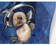
• An 18 year old girl visited the clinic with a chief complaint of intermittent pain from past 4 to 5 months .Medical History was non contributory. Clinical Examination showed deep occlusal caries with mandibular left first molar #36.

Figure 1: Mandibular left first molar [#36].

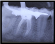
Figure 2: MMC also obturated.
• Pup sensibility showed no response to cold test as well as Eelectric Pulp Tester.
• Radiographic Evaluation showed Coronal radiolucency overlapping pulp space with Periodontal ligament space widening and periapical radiolucency. So diagnosis of pulpal necrosis with chronic apical periodontitis was given.
• Endodontic access cavity was performed under magnification using loops and middle mesial canal was found.
• Cleaning and shaping of all canals was done, which was followed by obturation of root canals.
Summary
There are numerous cases in the literature concerning the unusual anatomy of the mandibular first molar. The presence of a third canal in the mesial root of mandibular molars has been reported to have an incidence rate of 1 to 15%. This additional canal may be independent with a separate foramen, or the additional canal may have a separate foramen and join apically with either the mesiobuccal or mesiolingual canal. Instrumentation is one of the key factors in the success of endodontic therapy; therefore, the clinician should be aware of the incidence of these extra canals in the mandibular first molar. The clinician can then perform a thorough examination of the pulp chamber to insure complete debridement of all canals. This increases the chance for long-term successful endodontic therapy.
References
