
Case Report
Ann Surg Perioper Care. 2022; 7(1): 1052.
Osteochondral Lesions of the Talus (OLTs): 2 Case Reports
Zaizi A*, Badaoui R, Oussama A, Boukhris J, Chafry B, Benchebba D and Boussouga M
Department of Orthopaedic Surgery & Traumatology II, Mohamed V Military Hospital, Faculty of Medicine and Pharmacy, Mohamed V University, Morocco
*Corresponding author: Abderrahim Zaizi, Doctor at Department of Orthopaedic Surgery & Traumatology II, Mohamed V Military Hospital, Faculty of Medicine and Pharmacy, Mohamed V University, Rabat 10100, Morocco
Received: October 10, 2022; Accepted: November 07, 2022; Published: November 14, 2022
Abstract
Osteochondral Lesions of the Talus (OLTs) or (LODA) in French nomenclature are common and challenging conditions. They are typically correlated to ankle trauma; nevertheless, nontraumatic etiologies have been described. The diagnosis should be considered in the face of chronic ankle pain, particularly after a correctly treated sprain, and confirmed by CT-scan or MRI of the ankle.
We report two case reports treated arthroscopically with very good outcomes.
Keywords: Ankle; Osteochondral; Microfracture; Mosaicplasty
Introduction
Osteochondral Lesions of the Talus are rare lesions, which are mainly seen in young athletes, following ankle trauma in most of cases or nontraumatic [1]. Historically, multiple terms were used to term these lesions including osteochondritis dissecans, trans chondral talar fracture, and osteochondral talar fracture. However, it’s actually commonly named osteochondral lesions of the talus, a nomenclature that was introduced first by Kouvalchouk [2].
Improvements in imaging and arthroscopy allowed a better diagnostic and therapeutic approach to these lesions.
Case Reports
We report two patients suffering from Osteochondral Lesions of the Talus in our orthopedic trauma department between 2018 and 2022.
The first patient was a 27-year-old soldier, who had experienced a severe sprain of his left ankle, treated in another structure for 3 months, combining cast immobilization and functional rehabilitation but without improvement. Arrived at our consultation an MRI was requested, objectifying an antero-lateral talar osteochondral lesion (Figure 1).
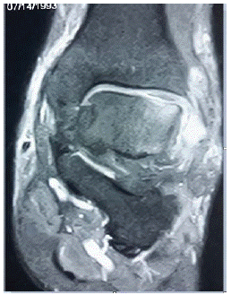
Figure 1: First case MRI showing antero-lateral talar osteochondral lesion.
The second patient was a 47-year-old woman without any history of trauma who complains of chronic mechanical pain in her right ankle, the radiological assessment was in favor of medial talar osteochondral lesion (Figure 2).
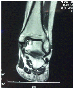
Figure 2: Second case MRI showing medial talar osteochondral lesion.
The two patients underwent arthroscopic treatment, in a supine position with a tourniquet at the root of the ipsilateral thigh without ankle traction (Figure 3). After exploration of the various anterior compartments of the ankle and visualization of the osteochondral lesion which was less of 1.5 cm for both patients, a decision of micro fracturing technique was made. Results were successively good and excellent according to the AOFAS score after 6 months follow-up.
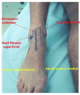
Figure 3: Landmarks of ankle anterior arthroscopic approaches.
Discussion
Osteochondral Lesions of the Talus (OLTs) are rare lesions which are mainly seen in young patient, with a clear male predominance [1,2]. Many hypotheses on the origin of OLTs were suggested, Traumatic context is involved frequently and preferentially located on the lateral side. Other non-traumatic causes exist and sit preferentially in the medial side of talus; therefore necrotic forms evoke vascular, synovial or metabolic etiologies with or without micro traumatic context or localized hyper pressure. In addition, non-traumatic OLTs can also be familial [3].
Concerning location of osteochondral lesion, medial ones are more frequent than lateral and differed in their characteristics, Thus, Medial lesions are profounder with extension into subchondral bone, and they often develop into cystic lesions. In the other side lateral osteochondral lesions, which are more commonly associated with a traumatic condition, are superficial and have predisposition to displacement. However, other study findings support the notion that the most common osteochondral lesions are not the conventionally described anterolateral and poster medial lesions, but rather central medial and central lateral lesions [4].
Symptoms are non-specific; patient can have pain, blocking, jumping, instability or joint swelling, and most often moderate limitation of joint mobility.
MRI is the examination of choice for the diagnosis of OLTs and detection of suspected lesions that are not seen on initial plain radiographs, as well as for the therapeutic decision because of very fine analysis of the bone matrix and the cartilaginous cap [5]. Several radiological classifications are used, since those of Berndt and Harty in 1959 based on radiographic findings in four stages. This classification system was later modified by Loomer and associates with the addition of stage V to describe lesions with a cystic component 16. Stage I defined a compression of subchondral bone, stage II designed partially detached osteochondral fragment, stage III completed etachment of osteochondral fragment remaining in fragment bed, stage IV displaced osteochondral fragment, finally stage V presence of a cystic component. In addition, Ferkel and Sgaglione’s classification define four types based on CT-scan. Stage I designed Cystic lesion within the dome of the talus, intact roof on all views. Stage IIA: cystic lesion with communication to the talar dome surface. Stage IIB: open articular surface lesion with overlying nondisplaced fragment. Stage III: nondisplaced lesion with lucency. Stage IV: displaced fragment [6,7].
Mitz classification based on MRI imaging is the preferred one, it defines five grades; grade 0 normal, grade I hyperintense but intact cartilage surface, grade II fibrillations of fissures not extending to bone, grade III flap present or exposed bone, grade IV loose, nondisplaced fragment, and grade V displaced fragment [8].
The treatment of these lesions remains in the majority of cases surgical, accessible to arthroscopic or conventional surgery aiming to remove loose fragment and create an environment amenable to fibro cartilaginous proliferation through micro fracturing or resurfacing with hyaline cartilage or osteochondral auto graft implantation using the Mosaicplasty or osteochondral allograft. Arthroscopic surgery is the treatment of choice particularly in posterior lesions, it is an effective, minimally invasive and safe procedure with low incidence of complications. In contrast, open approaches especially through malleolar osteotomies is at risk of nonunion or malunion and produce significant tissue trauma and may be associated with postoperative stiffness, prolonged rehabilitation time, and poor cosmetic appearance [9,10].
Conclusion
OLTs are rare condition that often goes unnoticed; the assessment must include standard x-rays in addition to CT-scan or more suitable MRI imaging. The surgical management of choice is ankle arthroscopy that gives simple follow-up and a rapid resumption of daily activities.
References
- Shimozono Y, Coale M, Yasui Y, O’Halloran A, Deyer TW, Kennedy JG. Subchondral bone degradation following microfracture for osteochondral lesions of the talus. Am J Sports Med. 2018; 46: 642-648.
- Kouvalchouk JF, Watin-Augouard L. Lésions ostéochondrales du dôme astragalien avec nécrose partielle. Rev Chir Orthop. 1990; 76: 480-9.
- Hangody L, Dobos J, Baló E, Pánics G, Hangody LR, Berkes I. Clinical experiences with autologous osteochondral mosaicplasty in an athletic population: a 17-year prospective multicenter study. Am J Sports Med. 2010; 38: 1125–33.
- Shimozono Y, Hurley ET, Myerson CL, Kennedy JG. Good clinical and functional outcomes at mid-term following autologous osteochondral transplantation for osteochondral lesions of the talus. Knee Surg Sports Traumatol Arthrosc. 2018; 26: 3055-62.
- Prado MP, Kennedy JG, Raduan F, Nery C. Diagnosis and treatment of osteochondral lesions of the ankle: current concepts. Rev Bras Ortop. 2016; 51: 489-500.
- R. Loomer, C. Fischer. Osteochondral lesions of the talus. Am J Sports Med. 1993; 21: 13-19.
- Ferkel RD, Zanotti RM, Komenda GA, Sgaglione NA, Cheng MS, Applegate GR et al. Arthroscopic treatment of chronicosteochondral lesions of the talus: long-term results. Am J Sports Med. 2008; 36: 1750–62.
- DN Mintz, GS Tashjian, Connell DA, Deland JT, O’Malley M, et al. Osteochondral lesions of the talus: a new magnetic resonance grading system with arthroscopic correlation. Arthroscopy. 2003; 19: 353-59.
- VanTienderen RJ, Dunn JC, Kusnezov N, Orr JD. Osteochondral Allograft Transfer for Treatment of Osteochondral Lesions of the Talus: A Systematic Review. Arthroscopy. 2017; 33: 217-22.
- de l’Escalopier N, Amouyel T, Mainard D, Lopes R, Cordier G, et al. Longterm outcome for repair of osteochondral lesions of the talus by osteochondral autograft: A series of 56 Mosaicplasties®. Orthop Traumatol Surg Res. 2021; 107: 103075.