Case Report
Ann Surg Perioper Care. 2025; 10(1): 1069.
Kidney Hydatid Cyst in Western Countries: Is Minimally Invasive Surgery Safe in Children?
Macchia I¹,², Cerchia E¹, Serpentino M¹,², Ruggero E¹,², Cirigliano L¹, Catti M¹ and Gerocarni Nappo S¹
1Pediatric Urology Unit, Department of Public Health and Pediatric Sciences, Regina Margherita Children’s Hospital, Turin, Italy
2Pediatric Surgery Unit, Department of Women’s and Children’s Health, University of Padua, Padua, Italy
*Corresponding author: Dr. Cerchia E, Pediatric Urology Unit, Department of Public Health and Pediatric Sciences, Regina Margherita Children’s Hospital, 10122, Turin, Italy Email: elisacerchia@gmail.com
Received: January 24, 2025; Accepted: February 05, 2025; Published: February 10, 2025
Abstract
Hydatid disease is a rural zoonotic parasitic infection. It predominantly affects liver and lungs, rarely kidneys (1.9% in children). The main treatment is surgery, either open or MIS (minimal invasive surgery). This is a case report of a 10-year-old boy who was found to have a hydatid cyst, located in the upper pole of the left kidney. The patient underwent left lower lobectomy for pulmonary cyst followed by laparoscopic transperitoneal excision of left renal cyst. Preoperative albendazole therapy was started to reduce the risk of cyst spillage and recurrence. This was continued for four weeks post-operatively. Complete excision of the cyst, preservation of the renal parenchyma and safe removal without contamination or anaphylaxis were achieved using a minimally invasive laparoscopic approach. The post-operative course was uneventful. Followup imaging at six months confirmed resolution without recurrence. Minimally invasive laparoscopic techniques, either transperitoneal or retroperitoneal, offer excellent exposure, reduced recovery time and minimised morbidity. This case highlights the safety and efficacy of laparoscopic management. It demonstrates its potential as the treatment of choice even in regions with limited experience in the management of echinococcosis.
Keywords: Hydatid cyst; Echinococcus; Laparoscopy; Pediatric urology; Minimally invasive surgery
Abbreviations
E: Echinococcus; CE: Cystic Echinococcosis; CT: Computed Tomography; MRI: Magnetic Resonance Imaging
Introduction
Hydatid disease or echinococcosis is a chronic parasitic disorder caused by the larval stage of Echinococcus (E) granulosus complex, E. multilocularis, E. vogeli or E. granolosus with humans as intermediate hosts [1]. This disease is a typical endemic zoonosis in rural areas with sheep breeding such as Asia, East Africa, Central Europe and some parts of South America [2]. Despite hydatid cyst may occur in any site of the human body due to haematogenous dissemination, the liver (55-75% in adults and 28% in children) and the lungs (18- 35% in adults and 64% in children) are the most commonly involved organs [3]. Only 10% of cases occur elsewhere, with the kidney being the third most common site (2-3% in adult and 1.9% in paediatric population) [4]. The cysts are more commonly detected in the 20-40 year age group but they often develop in childhood and subsequently slowly grow at a rate of 1-3 cm per year. The diagnosis is therefore often delayed because the cyst is asymptomatic for long periods. The clinical complains are variable and depend on the location, the size and the complication of the cyst. The most common symptom is abdominal pain, but the clinical features may be non-specific [5]. Renal hydatid cyst may mimic other renal pathologies and may present with haematuria, dilatation of the renal collecting system, flank pain and a palpable abdominal mass. Rupture of the cyst into the renal collecting system may result in renal colic and hydatiduria, while rupture of larger cysts may lead to a severe immune response [6]. Excision of the cyst with preservation of the renal parenchyma and avoiding contamination of the patient are the primary surgical goals in the treatment of renal hydatid cysts. Current practice lacks a solid consensus on surgical approach, but historically most authors have recommended open surgery. As an alternative, minimally invasive techniques have increasingly demonstrated their efficacy [7]. We present a case of renal echinococcosis in a 10 year old boy due to its rarity in childhood in a western country, and propose its treatment by a minimally invasive approach as a safe surgical option.
Case Presentation
A 10-year-old child (weight kg 30,8, height 140 cm) was admitted to hospital with fever and chest pain. A chest x-ray, abdominal ultrasound and thoraco-abdominal CT scan revealed a hypodense pulmonary lesion of fluid appearance with atelectasis of the adjacent lung parenchyma and a hypodense lesion of cystic appearance at the level of the upper pole of the ipsilateral kidney. Urinary symptoms were not reported. Blood and urine tests were normal. Throat swab, Quantiferon test, antibodies for Entamoeba and Echinococcus were negative. Stool and gastric aspirate pathogen tests were also negative. Initial surgery of the lung lesion was planned after a multidisciplinary assessment of the case with involvement of infectious disease colleagues. Given the uncertain diagnosis, the first surgical procedure was a left lower lobectomy performed through thoracotomy. The postoperative course was uneventful. Histological examination was compatible with an Echinococcus cyst. Treatment of the renal cyst was considered one month later in view of the histological examination and the exponential growth of the renal cyst lesion. The preoperative abdominal MRI confirmed the presence of an extensive renal cystic lesion in the upper pole of the left kidney, sized 55x60x60 mm, with homogeneous hyperintense T2 and hypointense T1 signal without enhancement, in close proximity to the splenic hilum, the tail of the pancreas, the adrenal gland and the upper renal calyxes. Treatment with albendazole (400 mg twice daily) was therefore started four days before surgery. A minimally invasive laparoscopic transperitoneal approach was planned. The technical details of the procedure are explained as follows.
Surgical Technique
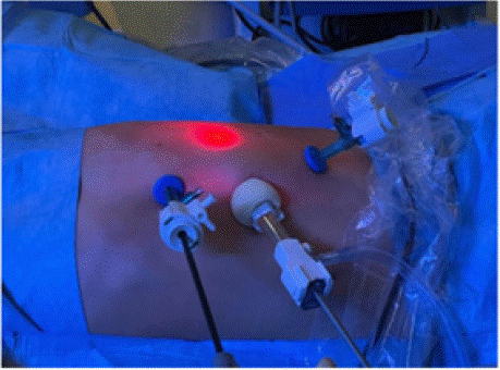
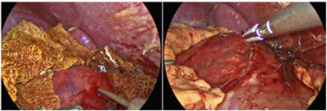
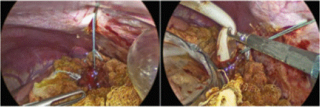
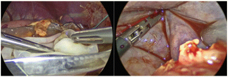
The patient was placed in a supine position with a 45° subcostal tilt under general anesthesia. Four trocars were placed a 12-mm umbilical trocar (for a 5-mm 30° optic), two 5-mm trocars in the epigastric and left iliac fossa and a 10-mm trocar in the left flank (Figure 1). A Ligasure device was used to incise along Toldt’s line, medializing the descending colon. The Gerota fascia was opened, revealing significant distortion of the upper kidney profile without clear definition of hydatid cyst from renal parenchyma and clear demarcation from adrenal gland. Betadine-soaked gauzes, irrigated with 30% hypertonic saline solution, were placed around the cyst. A laparoscopic needle was used to puncture the cyst and aspirate 120 ml of rock water-like fluid, which was sent for cytological examination (Figure 2). The cyst was then filled with 100 ml of hypertonic solution (30% saline), left in place for 15 minutes, and subsequently aspirated without spillage. To allow stable and safe evacuation of the cyst contents, a tranparietal traction suture was placed on the roof of the cyst. An endobag was inserted and opened next to the cyst. Using a suction device, the intact germinal membrane was aspirated (Figure 3) and placed in the endobag without contaminating nearby tissues. No evidence of daughter cysts was found in the remaining cavity, which was inspected with the optic and washed with hypertonic solution (Figure 4. Through the umbilical incision, the endobag and its contents were removed intact. Betadine soaked gauze was removed in a second endobag. A 15 Ch drain was placed in the cyst cavity after a partial resection of the wall of the cyst and the placement of the omentum in the remaining cavity. Both the surgical procedure and the postoperative period were uneventful. The abdominal drain was silent removed on postoperative day 2 and patient was discharged on postoperative day 4. Albendazole was continued for four weeks postoperatively. Histological examination revealed fibrosclerotic tissue fragments with an intense eosinophilic granulocytic inflammation and a multi-layered lamellar chitinous fragment. Bacterioscopy, parasitology and cyst culture were negative. Clinical recovery was excellent. Initial postoperative ultrasound showed a heterogeneously hyperechoic area with irregular margins at the upper pole of the left kidney at the surgical site, which resolved at subsequent examinations. At 3 and 6 months, no further hydatid cysts were found at abdominal ultrasound and brain MRI, and currently the patient is symptom-free.

    Figure 1: Laparoscopic approach for hydatid cyst sided at the upper pole of
left kidney. View of trocars position.

Figure 2: Laparoscopic aspiration of renal hydatid cyst.

    Figure 3: After placement of a transparietal stitch to stabilise the cyst,
laparoscopic removal of the removal of germinating membrane.

    Figure 4: The germinating membrane was placed in the endobag and no
evidence of daughter cysts was found in the remaining cavity.
Discussion
Hydatid disease is a major public health problem in many countries and requires epidemiological control measures for its eradication. Accidental ingestion of E. granulosus embryonated eggs after close contact with dogs usually causes this disease. Although the vast majority of the reported cases of cystic Echinococcus are in adults, most human infections occur during childhood and adolescence. Many series describe the predominance of boys over girls, which could be attributed to behavioural differences between the two sexes with boys having more outdoor exposure, as suggested by Tantawy et al [8]. The age of patients varies from 1 to14 years, although hydatid cysts have also been reported in a 6-month-old infant [9]. Our patient was a 10-year-old male child, in line with the literature. Hydatidosis may affect the brain, heart, kidneys, ureters, spleen, uterus, fallopian tubes, pancreas, diaphragm, bones and muscles [10]. Mamishi et al [11] retrospectively reviewed 31 children and reported multiorgan involvement only in three cases. The two most common sites of involvement are the lungs and liver, although no organ is spared. The hydatid cyst may be unilocular or multiloculated and thin or thick walled. The natural history of an untreated cyst may be calcification and death, but more often there is gradual but progressive enlargement of the cyst. An abdominal mass is the most common complaint in cases of liver involvement. Intraperitoneal rupture of a hydatid cyst is a life-threatening emergency that can be fatal [9], as the cyst fluid is highly allergenic and carries a risk of anaphylaxis. Renal involvement is extremely rare, accounting for only 1.9% of paediatric cases, and isolated renal involvement is even a rarer condition. Because of the low incidence, most reports of renal hydatid disease are case reports, and is therefore difficult to draw epidemiological data. In adult literature it is reported a predominance for left kidney localisation, Imani F et al reported 10 cases of renal CE of which 8 in the left side, as them also Gögüs Ç et al reported 20 cases of which 14 in the left side and Huang M. et al reported 18 patients of which 12 in the left side hypotizing that it could be due to the shorter left renal artery [12]. In the paediatric literature, both sides seem equally affected as in Table 1. In our case both renal and pulmonary cysts were left sided. The majority of renal hydatid cysts are asymptomatic in the early stages, but if the cysts communicate with the renal collectic system, hematuria or pyelocaliceal dilation can occurr. Symptomatic cases present with flank pain and a palpable mass. In some instances, rupture of large cysts may trigger severe immune responses, including urticaria or even anaphylaxis. Additionally, rupture into the renal collecting system may result in renal colic and hydatiduria. In our case, the diagnosis was incidental and made after the appearance of fever and chest pain related to the left thoracic cystic site, while the patient did not have urinary symptoms or a palpable abdominal mass. Imaging plays a key role in the diagnosis and staging of hydatid disease, whereas serology has only a minor confirmatory role due to the high rate of false negative results. Ultrasonography is the most important tool in hydatid disease and clearly demonstrates the floating membranes, daughter cysts and hydatid sand feature characteristic of pure cystic lesions. The World Health Organisation Informal Working Group (WHO-IWGE) has developed an ultrasound classification for staging the disease [13]:CE 1: unilocular fluid collection/single cyst with double line sign or "snowflake sign".
    Year
    Autors
    Country
    Number of patients
    Side
    Surgery


    2025
    Macchia et al
    Italy
    1
    Left
    Laparoscopic Cystectomy


    2022
    Aggarwal et al
    India
    1
    Left
    Laparoscopic    Cystectomy


    2020
    Kara et    al
    Turkey
    1
    Left
    Laparoscopic partial nephrectomy


    2016
    Uçar et al
    Turkey
    1
    Right
    Laparoscopic    Cystectomy with diode laser


    2013
    Ksiaa et al
    Tunisia
    4
    3 Right/1    Left
    3    Laparoscopic Cystectomy/ 1 Retroperitoneal Cystectomy


    2013
    De    Carli  et al
    Argentina
    1
    Left
    Retroperitoneal    Cystectomy


    2010
    Divarci et al
    Turkey
    1
    Right
    Laparoscopic    Cystectomy


    2008
    Onal et al
    Turkey
    1
    Right
    Laparoscopic    Cystectomy


    2005
     Bilen    et al
    Turkey
    1
    Right
    Laparoscopic    Cystectomy
Table 1: Literature review data for laparoscopic treatment of renal hydatid cysts in the pediatric population.
• CE 2: a) multivesicular/multiseptate cyst partially or completely filling the unilocular mother cyst, b) cyst septations as "wheel-like structures", c) contained daughter cyst as "honeycomb" or "rosette-like".
• CE 3A: a) fluid collection with detached membrane (waterlily sign), b) presence of daughter cysts in a solid matrix.
• CE 4: Cysts with heterogeneous hypoechoic/hyperechoic matrix without daughter cysts.
• CE 5: solid cystic wall.
CE1 and CE2 indicate active disease. CE3 indicates a transitional stage where the cyst is weakened, while CE4 and CE5 indicate inactive disease. In our clinical case, the renal hydatid cyst corresponded to a stage 2a in the classification and was therefore an active renal hydatid cyst. CT scan is superior to other imaging modalities in the observation of intracystic gas and minute calcifications, and in anatomical mapping cysts can be identified as single or multiple, and uni- or multilocular. Medical management, percutaneous procedures and surgical intervention using open or minimally invasive approaches are all possible strategies available for the treatment of renal hydatid cysts. Percutaneous management has been reported as a safe option (PAIR puncture, aspiration, injection of scolicidal agent, re-aspiration) under ultrasonography guidance as an alternative treatment for renal hydatid cysts not involving the collecting system [14]. Disadvantages include the risk of spreading daughter cysts and fatal anaphylaxis. Surgery is the treatment of choice for renal hydatid cysts for either resolution of the disease, symptom relief and sometimes diagnosis. Whenever possible, renal-sparing surgery must be pursued. Simple excision of the cyst is the preferred procedure but does not completely prevent recurrence. Nephrectomy is required if the kidney is compromised. In recent years minimally invasive treatment of renal hydatid cyst has been proposed in children. Bilen CY et al [15] first reported transperitoneal treatment of a huge renal cyst of the upper pole of the right kidney with a diameter of 15 cm in a 13-year boy, the procedure lasting 4.5 hours was performed laparoscopically and was complicated by urinary leakage which was managed with JJ stent placement. Preserved renal function and no recurrence was noted on CT at follow-up. Laparoscopic management of a right renal cyst in a 5-year-old boy was also reported by Onal B et al with no complications [16]. Ksiaa A et al [17] published a small series of 4 paediatric cases between the ages of 7 and 14 years who were treated with minimally invasive surgery via a transperitoneal approach or a retroperitoneal approach. In the presence of a liver cyst, the transperitoneal approach was chosen. Two other authors De Carli C et al, Divarci E et al [18] reported minimally invasive treatment of a renal cyst in a 10-year-old and a 17-year-old patient using retroperitoneoscopic approaches (Table 1). The transperitoneal route provides a larger working space, but spilling the cyst contents is particularly feared, thus risking anaphylaxis. To prevent this, the cyst is packed tightly with gauze soaked in 30% saline or other scolicidal solutions containing 1% iodine, 10% cetrimide, 10% povidone-iodine or 0.5% silver nitrate. Only one case of anaphylactic shock during laparoscopy for hepatic hydatid cyst was reported in the 1990s. Bilen et al showed that laparoscopy, when performed correctly, does not increase the risk of spillage and that pneumoperitoneum protects against spillage [19]. In our case, due to the location of the cyst at the upper pole of the left kidney, we opted for a transperitoneal approach. All possible precautions were taken to avoid spilling the contents into the peritoneum, which could lead to recurrence, spread or development of severe anaphylactic shock. Other advantages of the laparoscopic approach were the ability to insert the camera into the cyst cavity for inspection and to ensure that no daughter cysts or lamina propria were missed, as described by the previous author [20]. The medical management of renal hydatidosis is a long way from being a realistic alternative to surgery and should only be considered as an adjuvant therapy. Albendazole for one week to one month before surgery may reduce intraoperative tension of the echinococcal cyst, prevent spread of echinococcus during puncture, and may kill or reduce activity of echinococcal larvae. Continued use of albendazole for up to 3 months after surgery may also reduce the incidence of postoperative recurrence, particularly if the cystic fluid has spread during the operation. In renal hydatid cysts, the efficacy of this treatment has not been proven, and these drugs are mainly used prophylactically before and after surgery to prevent the cyst from spreading. In our case, we administered oral albendazole 400mg twice daily for 4 days preoperatively and for 4 weeks postoperatively.
Conclusions
A renal hydatic cyst in a child is a very rare occurrence. Multimodal management with medical therapy in combination with nephronsparing surgery is the treatment of choice. Minimally invasive surgery through either a transperitoneal or retroperitoneal approach can be a safe and successful option for the treatment of a renal hydatid cyst. It provides good exposure of the cyst, an adequate level of safety, drainage of the cyst and complete removal of the germinal membrane without spillage, inspection of the cyst with the telescope, reduced pain and rapid recovery. We therefore suggest that it should be considered as the treatment of choice not only in countries where Echinococcus is endemic, but also in Western countries with limited experience of this disease, even in these atypical presentations.
References
- Almulhim, Abdulaziz M and Savio John. “Echinococcus Granulosus (Hydatid Cysts, Echinococcosis).” PubMed, StatPearlsPublishing. 2020.
- Lodhia J, Chugulu S, Sadiq A, Msuya D, Mremi A. Giant isolated hydatid lung cyst: Two case reports. J. Med. Case Rep. 2020; 14: 200.
- Patil RT, Parelkar SV & Sanghvi B. Laparoscopic management of renal and hepatic hydatidosis in a child: a case report and review of literature. International Surgery Journal. 2017; 4: 1793–1796.
- Ines M, Mariem BL, Marwa M, Amina BS, Chiraz H. Isolated breast hydatid cyst: Imaging features. Clin. Case Rep. 2022; 10: e06362.
- Paduraru AA, Lupu MA, Popoiu CM, Stanciulescu MC, Tirnea L, Boia ES, et al. Cystic Echinococcosis in Hospitalized Children from Western Romania: A 25-Year Retrospective Study. Biomedicines. 2024; 12: 281.
- Hamidi Madani A, Enshaei A, Pourreza F, Esmaeili S, Hamidi Madani M. Macroscopic Hydatiduria: An Uncommon Pathognomonic Presentation of Renal Hydatid Disease. Iran J Public Health. 2015; 44: 1283-1287.
- Rexiati M, Mutalifu A, Azhati B, Wang W, Yang H, Sheyhedin I, Wang Y. Diagnosis and surgical treatment of renal hydatid disease: a retrospective analysis of 30 cases. PLoS One. 2014; 9: e96602.
- Tantawy, Ismail M. “Hydatid Cysts in Children.” Annals of Pediatric Surgery. 2010; 6: 98-104.
- Jairajpuri ZS, Jetley S, Hassan MJ, Hussain, M. Hydatid disease in childhood: revisited report of an interesting case. Journal of parasitic diseases. 2012; 36: 265-268.
- Abu-Eshy SA. “Some rare presentations of hydatid cyst (Echinococcus granulosus).” Journal of the Royal College of Surgeons of Edinburgh. 1998; 43: 347-352
- Mamishi S, Sagheb S, Pourakbari B. Hydatid disease in Iranian children. Journal of Microbiology, Immunology, and Infection = Wei Mian yu gan ran za zhi. 2007; 40: 428-431.
- Mirlohi SH, Tajfirooz S, Raji H, Akhavan S. Coexistence of kidney and lung hydatid cyst in a child: A case report. Respir Med Case Rep. 2024; 52: 102138.
- WHO Informal Working Group. International classification of ultrasound images in cystic echinococcosis for application in clinical and field epidemiological settings. Acta Trop. 2003; 85: 253-261.
- Cretu CM, Codreanu RR, Mastalier B, Popa LG, Cordos I, et al. Albendazole associated to surgery or minimally invasive procedures for hydatid disease– how much and how long. Chirurgia (Bucur). 2012; 107: 15–21.
- Bilen CY, Ozkaya O, Sarikaya S, Asçi R & Büyükalpelli R. Laparoscopic excision of renal hydatid cyst in a preadolescent. Journal of pediatric urology. 2006; 2: 210-213.
- Onal B, Demirkesen O, Citgez S, Argun B, Oner A. Laparoscopic treatment of unilocular renal hydatid cyst mimicking a simple cyst in a child. J Pediatr Urol. 2008; 4: 477-479.
- Ksiaa A, Zitouni H, Zrig A, Kerkeni Y, Sahnoun L, Chahed J, et al. Videoassisted surgery in the management of hydatid renal cyst in children. J Pediatr Surg. 2013; 48: E17-19.
- De Carli C, Viale A, Perez Lau F, Campaña R. Retroperitoneal laparoscopic approach for renal hydatid cyst in children. A technical report. J Pediatr Urol. 2013; 9: e35-38.
- Bilen CY, Ozkaya O, Sarikaya S, Asci R, Büyükalpelli. Laparoscopic excision of renal hydatid cyst in a preadolescent. Journal of pediatric urology. 2006; 2: 210-213.
- Patil RT, Parelkar SV, Sanghvi B. Laparoscopic management of renal and hepatic hydatidosis in a child: a case report and review of literature. Int Surg J [Internet]. 2017; 4: 1793-1796.