
Research Article
Austin J Anat. 2016; 3(3): 1056.
Laparoscopic Anatomy of the Abdomen in Dorsal Recumbent Male Donkey
Mohamed NA¹* and Wefky AM²
¹Department of Anatomy, Faculty of Veterinary Medicine, Assiut University, Egypt
²Department of Surgery, Faculty of Veterinary Medicine, Assiut University, Egypt
*Corresponding author: Mohamed NA, Department of Anatomy, Faculty of Veterinary Medicine, Assiut University, New Valley, Egypt
Received: September 20, 2016; Accepted: October 17, 2016; Published: October 17, 2016;
Abstract
Laparoscopic anatomy of the abdomen in dorsal recumbent male donkey was an applied anatomical study. Provide an accurate and detailed description of the laparoscopic anatomy of the abdomen of donkey positioned in dorsal recumbency was considered a diagnostic and a therapeutic surgical importance. The laparoscopy was performed on ten adult apparently healthy male donkeys in average of 7-12 years old, in order to record the normal laparoscopic anatomy of abdomen in dorsal recumbency. The preoperative techniques were considered. General anesthesia was induced and maintained with total intravenous agents. Laparoscope was delivered into the abdomen via an umbilical main cannula. Laparoscope and associated video recording system were used to obtain a clear, magnified and panoramic shoots of the abdominal cavity. The anatomical findings were more real and vital than that of fixed condition. The prestaltic motility as well as the vascularization of the serous layer of peritoneum and the abdominal organs was notified. The study was fully described two dangerous triangular areas; TD and TP especially during endoscopic repair of inguinal hernia. The work considered as a gate to further endoscopic anatomy and the donkey was an ideal experimental model.
Keywords: Donkey; Laparoscopy; Dorsal recumbency; Laparoscopic anatomy
Introduction
Endoscopic anatomy of live animal differs than structural anatomy in cadaver. For successful laparoscopy, surgeons need to gain full detailed anatomical information about structures dealing within the abdomen. Different studies described the laparoscopic anatomy of dogs, Atiba [1] and equines, Galuppo et al. [2]. Most of the authors deal with describing fixed animal bodies using formalin 10% concentrate El- Hagri [3], Nickle et al. [4], Dyce et al. [5] & Konig and Liebichin [6] in domestic animals.
In the recent study, try to describe the abdominal structures in the life state. The anatomical describing terminology according to NAV [7].
Laparoscopy is an endoscopic surgical procedure by which a variety of intra-abdominal and pelvic diagnostic and therapeutic procedures can be performed Kumar [8].
Since the first canine experimental laparoscopic procedure in 1901, laparoscopic procedures were described in different animal species including equine Hendrickson [9], cattle, Babkine and Desrochers [10], small ruminants Dovenski et al. [11], canines Abd El-Alim [12] and in some exotic animals Anderson et al. [13]. Laparoscopy is a minimal invasive procedure aims to achieve the purpose of traditional surgical procedures with minimal intra and post-operative complications and as well as tissue damage.
Food withholding period, degree of pneumoperitoneum and animal position are main factors affecting organ arrangement and relationship within the abdominal cavity, another factor affecting the endoscopic view of abdominal cavity is the endoscope port position.
Materials and Methods
Ten adult apparently healthy local breed donkeys with average weight 230-250 kgs aged from 7-12 years were submitted for the study.
Animals were fasted for 24 hours and water was allowed ad lib. Prior surgery, general health chick up was performed to ascertain good general health status of experimental animals. Antibiotics and anti-inflammatories were administered 12 hours prior and 72 hours post surgery.
Animals were sedated by using xylazine Hcl in a dose of 1.1 mg/kg Bwt and anesthesia was induced by Ketamine Hcl in a dose of 2.2 mg/kg Bwt Anesthesia was maintained by repeated one third ketamine dose Delling [14]. Local analgesic was infiltrated at the umbilicus. Animals were dorsally positioned and secured by robes to the surgical table.
Ventral abdomen was clipped, scrubbed, draped and prepared for aseptic surgery. The umbilicus incised and grasped by two towel clamps and the main portal safety cannula inserted directly into the abdominal cavity [15].
Standard pneumoperitoneum to 12 mm hg was created using automatic insufflator. Rigid endoscope with 10 mm outer diameter, 45 cm length and zero degree viewing angles connected to high definition video camera and display were used to obtain a highly magnified intra abdominal view.
Xenon cold day light was used to illuminate the abdominal cavity. Anatomical structures of the abdominal cavity were obtained while animal in Trendelenburg position [the head tilted down the level of hind quarter] while the anatomy of cranial portion of abdominal cavity was obtained with the animal positioned in reverse Trendelenburg position [the head up position] (Figure 1).
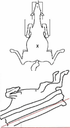
    Figure 1:  Diagram showing the position of examined donkey in dorsal
recumbency and (X) is the site of operation.
Anatomical features and organ arrangement and relations were recorded. The laparoscope removed and gas was permit to escape. Umbilical wound was sutured and antiseptics applied. Animals positioned in lateral recumbency and allowed for recovery (Figure 1).
Results
Laparoscopic investigation of the abdominal cavity in the dorsal recumbent male donkeys, reveals most of the anatomical structures and organs that lodged in the intrathoracic and intrapelvic portions of the abdominal cavity. Some of the internal organs of the abdomin may not appear in regard to the huge sized large intestinal mass. In general consideration, the anatomical examination in the live animals differs than that of dead fixed one. Although in the former condition, the investigator can examine the color of the peritoneum with the configuration of its vascularization as well as that of the organs and prestaltic movement of intestinal tract.
In both parts of the abdominal cavity; the cranial Intrathoracic and caudal Intrapelvic part: the large intestine especially the ventral parts of the ascending and descending colon as well as the jejunum of small intestine, form the disappearance of some abdominal organs.
Intestine (Figures 2-6&8)
It should be noted that the motile activity of live intestine in the examined cases reveals that the anatomical arrangement of the large and small parts of intestine is changed during examination in addition to the attended posture of investigation may shift the cecum to unseen area. The ventral ascending colon (colon ascendens) has right and left portions (pars dextrum & sinistrum); the former is related to the ventral parts of live. While the latter one is related to the spleen. These structures are connected together cranially by the sternal flexure (flexura sternalis).
The intestinal mass is similar in shape and they have an external segmentation, which are connected externally together by longitudinal fibrous bands (Taeniae coli) and these segmentations are known by haustra coli or sacculations. The descending colon (colon descendens) is lodged caudally at the pelvic inlet and characterized by double taeniae as well as two rows of sacculations that model the final form of fecal matter.
Regarding to the intrathoracic part of the abdominal cavity, the organs that significantly investigated are the caudoventral part of diaphragm, liver and spleen.
Diaphragma (Figures 2,3&4)
It should be mentioned that according to the extension of the ascending colon, which covers the dorsal parts and roots of diaphragm. Although, the laparoscopic examination in recumbent position detects the ventral parts of diaphragm; the fleshy parts (costal and sternal) (Pars costalis & pars sternalis) and central tendinous part (Centrum tendineum). The costal parts are attached to ribs laterally and met ventrally to form the sternal part on the xiphoid cartilage. Both halves of the costal part of diaphragm are broad flat muscle fibers measures about 10-13 cm in width, extends from the ribs laterally to the central tendinous. The later is characterized by its whitish fibrous appearance and branched phrenic veins in between. Both parts of diaphragm are covered by serous colorless parietal layer of peritoneum.
Hepar (Figures 3,4&5)
The cranioventrally exposed parts of liver, anatomically is reddish to brownish in color with sharp, tapered and pointed cranioventral border. The interlobar fissure (Icisura interlobares) which between the quadrate and left central lobes are characterized by the emergenceof the falciform and round ligament of liver (Lig. Teres hepatis), which pass ventrally on the abdominal wall to the umbilicus (Figure 2). The caudodorsal border of the liver was attached to the central tendenous part of diaphragm by the triangular ligament (Figure 4).
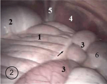
    Figure 2:  Photograph showing the cranial aspect of the abdominal cavity.
1. Colon ascendens pars ventrale dexterum
2. Colon ascendens pars dorsale dexterum
3. Jejunum
4. Diaphragma pars costalis
5. Falciform and lig, Teres hepatis
6. Colon ascendens pars ventrale sinistrum
The arrow indicates the taenia on the 1.
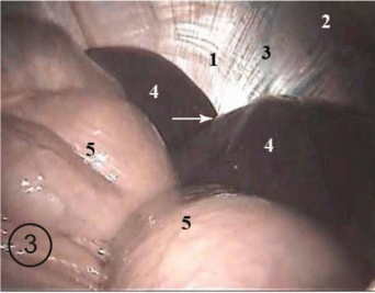
    Figure 3:  Photograph showing the cranial aspect of the abdominal cavity.
1. Diaphragma pars centrum tendineum
2. Diaphragma pars costalis
3. V. phrenici
4. Lobus hepatis sinister
5. Colon ascendens
6. (flexura sternalis) (diaphragmatica ventralis)
The arrow indicates the incisura interlobares hepar.
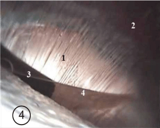
    Figure 4:  Photograph showing the cranial aspect of the abdominal cavity.
1. Diaphragma pars centrum tendineum.
2. Diaphragma pars costalis 3-Lobus hapatis sinister (margo dorsalis) 4- Left
triangular ligament of liver.
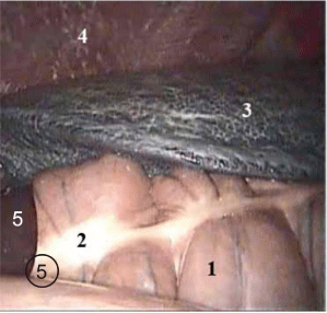
    Figure 5:  Photograph showing the left aspect of the intrathoracic part of the
abdominal cavity.
1. Colon ascendens pars ventrale sinistrum
2. Taenia
3. Lien, facies parietalis
4. Diaphragma pars costalis 5-hepar
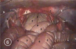
    Figure 6:  Photograph showing the caudal part of the abdominal cavity.
1. Lig. Inguinale 2-Distended vesica urinaria 3- Lig. Vesicae medianum
4-Vaginal ring 5-Haustrae colon descendens 6-Taenia 7-Ductus deferens
8-A &V. epigastrica caudalis profundus.
Lien (Figure 5)
The spleen is a bluish in color present in the left hypochondric subregion of the intrathoracic part of the abdominal cavity. It has two surfaces and borders. There are parietal and visceral surfaces the former is related to the lateral abdominal wall and the visceral one to the ascending colon.
The most important organ in the intrapelvic part of the abdominal cavity is the urinary bladder. It covers the overlaying structures; urogenital fold and rectum.
Vesica urinaria (Figures 6,7&8)
The urinary bladder is a distended or an empty sac that lies on the pubic tubercle and extends on the pubic symphysis. The anterior portion of the bladder is covered by serous parietal peritoneum while the posterior one is retroperitoneal. It presents between the vesico pubic pelvic pouches ventrally and genitovesical pouch dorsally. It is connected to the pelvic and abdominal wall laterally by a peritoneal fold, the right and left lateral vesico umbilical ligaments (Lig. Vesicae laterale). Each free border of the ligaments has a remnant of the right and left umbilical artery respectively which are the round ligaments of urinary bladder (Lig. Teres vesicae).
The cranioventral border of the bladder is connected to the floor of the abdominal cavity with a peritoneal fold; middle vesicoumbilical ligament (Lig. Vesicae medianum). The midpoint of junction between the folds of the bladder represented by the remnant of uracus that of embryo.
Peritoneum (Figures 7&9)
It is a color-less glistening vascularized serous membrane lines the abdominal wall internally. It has two portions; parietal and visceral part. The former is lined to the abdominal wall and covers the anterior portion of the organs of the pelvic cavity to form the pelvic pouches, in the recumbent poisoned cases the vesicopubic pouch is clearly seen. While the visceral one covers the abdominal organs and in some locations it forms a ligamentous attachment between the adjacent organs or fix the organs with the abdominal wall. The peritoneum carries minute capillary tree extends along the membrane.
The inguinal region is an important region from the anatomical point of view. The most characteristic anatomical structure is the spermatic cord which has vascular and nonvascular parts as well as mesentery. The former represented in the testicular artery (A.testicularis) and vein (V.testicularis) that enter and leave the testis respectively (Figure 9). While the non vascular part is the vas deferens which emerges from the cord to the pelvic urethra (Figure 8). The mesentery that lodged in between is the mesorchium which suspends the testis with the dorsal abdominal wall in the sublumbar region. All above mentioned structures reach and leave the testis via the vaginal ring.
It was an important to be notified that the investigator must be differentiated between the vaginal ring and deep inguinal ring. The vaginal ring is a slit like opening measures about 2-2.5 cm in width, which located on the deep inguinal ring on the caudal border of the internal abdominal obliqueus muscle (M. obliquus internus abdominis). This ring is formed due to invagination of the testicle with the vas deferens and surrounded blood vessels and bounded by the parietal peritoneum.
Ductus deferens (Figures 6,7,8,9&10)
The vas deferens emerges from the vaginal ring to the abdominal cavity and passes in a caudo ventromedial direction crossing the round ligament to cross caudo dorsally to the bladder. It should be attended that the triangular area that restricted laterally by the spermatic vessels and medially by the vas deferens, the parietal peritoneum covers on the external iliac artery and vein (A &V iliaca externa) as well as the obtiurator nerve (N. Obtiuratorius) which are pass within the area, so that it is a contra indicated to surgeons to do surgical interference in this area avoiding injury of that vital structures. This area known by the Triangle of Doom (TD). On the lateral side to TD, the imaginary iliopubic line encloses with the spermatic cord, a Triangular area of Pain (TP) that includes the femoral nerve (N. femoralis) and lateral cutaneous femoral n. (N. cutaneus femoris lateralis). The surgical activity is dangerous there. (Figures 7,8,9&10).
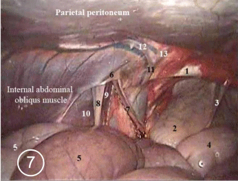
    Figure 7:  Photograph showing the triangle of doom.
1. Lig. inguinale
2. Distended vesica urinaria
3. Lig. Vesicae medianum
4. Colon descendens
5. Jejunum
6. Vaginal ring
7. Ductus deferens
8. Mesorchium
9. Testicular vessels
10. Cremastricus externus muscle
11. Truncus pudendoepigastricus
The dotted lines indicates triangle of doom.
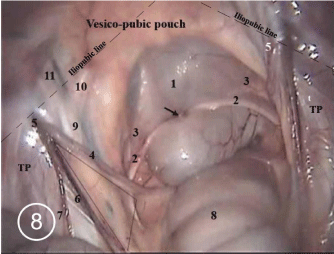
    Figure 8:  Photograph showing the pelvic inlet.
1. Distended vesica urinaria
2. Ligamentum teres vesicae
3. Lig. Vesicae laterale
4. Ductus deferens
5. Vaginal ring
6. V. testicularis
7. Colon descendens
8. Truncus pudendoepigastricus
9. V. epigastrica caudalis profundus
The arrow indicates the remnant of urachus.
The triangular area indicates the triangle of doom.
TP indicates the triangle of pain.
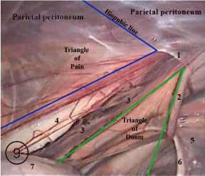
    Figure 9:  Photograph showing the inguinal region.
1. Vaginal ring
2. Ductus deferens
3. Mesorchium
4. Lig. Vesicae laterale
5. Ligamentum teres vesicae
6. Colon descendens
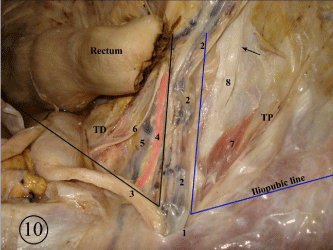
    Figure 10:  Photograph showing the inguinal region (Carcass dissection).
1. Vaginal ring
2. Testicularis
3. Ductus deferens
4. V. Iliaca externa
5. N. Obtiuratorius
6. M. cremaster
7. N. Femoralis
The arrow indicates N. cutaneus femoris lateralis.
TP within the bluish arms indicates the triangle of pain.
TD within the black arms indicates the triangle of doom.
Discussion
The endoscopic anatomical study in the living donkey of the recent work revealed that the most anatomical structures of the abdominal cavity in a recumbent position. The study notified the abdominal contents including the shape and the coloration of the serous peritoneum as well as the intestine with the adjacent organs. The anatomical findings were more real and vital than that of postmortem one and gave an accurate anatomical data for the surgeons. The result, which were more practical than that of most of anatomists.
The current descriptive findings recorded the motility of the intestine and absence of organ impressions as well as detecting real coloration of tissues. On the other hand, and according to the fixation process that depends up on replacing the blood by the formalin 10%, the authors described the organs in fixed state and preserved in the body in situ with direct contact together showing the impressions on the organs, in addition to discoloration of tissues and organs.
The laparoscopic appearance of the large intestine in this study notified disappearance of the cecum on the floor of the abdomen that is due to the preparation protocol before the endoscopy in addition to prestaltic motility. While Nickle et al. [4], Dyce et al. [5] & Konig and Liebich [6] in domestic animals, mentioned that the cecum ran on the floor of the abdominal cavity, it extended from the right paralumbar fossa caudally to the xiphoid region cranially.
Concerning the liver and spleen, the laparoscopic finding recorded them without impressions from adjacent organs as well as normal colorization. However, Nickle et al. [4] & Dyce et al. [5] in domestic animals cited impressions on both.
The anatomical structures of the diaphragm were similar with that of most of anatomists. El- Hagri, [3], Nickle et al. [4] & Dyce et al. [5] in domestic animals.
The laparoscopic anatomy of the serous peritoneum, showed in our study fullness of the capillaries with blood and vascularization of the membrane was clear. These statement were not recorded in the available literatures
The study is totally agreement with the authors in describing the urinary bladder. Nickle et al. [4], Dyce et al. [5] & Konig and Liebich [6] in domestic animals.
In the field of surgical anatomy, concerning the inguinal region was an important point of view, it should be attended that the testicular vessels and vas deferens comprises a triangular area known as triangle of doom; through it the external iliac vessels pass and the peritoneum covers up on, so it should be notified that it was dangerous to interfere or dissect within the area during the endoscopic repair of inguinal hernia, it could be fatal. A result which were totally agreement with that cited by Spaw [16]. While the recent findings added the presence of the obtiurator nerve within the area.
It was significant to point out the triangular area which are located laterally to the triangle of doom between the testicular vessels and the imagery iliopubic line, was a dangerous area, it contains the femoral nerve, lateral cutaneous femoral branch of femoral nerve, so the activity within the area may causing severe pain, the area named triangle of pain. Similar results were recorded by Spaw [16]. The described areas of doom and pain of the present study were scantly in the available veterinary anatomical literatures.
On the other hand, it is important to differentiate between the vaginal ring and the deep inguinal ring, as met with the opinion of Sisson, Grossman & Getty [17] in domestic animals.
Laparoscopy involves insertion of a rigid endoscope into the abdominal cavity through a minute abdominal incision which is usually the umbilicus of the patient; it provides the operator with a highly magnified image of the abdominal organs. In recent years laparoscopy had evolved almost all fields of traditional surgery, a complete thorough understanding of laparoscopic abdominal and pelvic anatomy of dorsal recumbent donkeys will enhance the diagnostic and therapeutic abilities of laparoscopy procedures.
In the present applied laparoscopic anatomical study of the abdominal cavity of dorsally recumbent donkeys, the mean procedure time was 35±7 minutes that permit thorough investigation of both cranial and caudal portions of the abdomen.
The total intravenous anesthetic protocol presented by Mama [18], was satisfying and provided an adequate anesthetic duration and proper efficacy which could be considered in short duration procedures with reduced intrathoracic compression and adequate intestinal evacuation which are recommended by El-Khamary et al. [19], while the standard laparoscopic procedures in dorsally recumbent animals are recommended to be performed under the effect of general inhalant anesthesia according to Fischer [20].
In order to avoid the heamodynamic hazards arising from pneumoperitoneum and putting the animal in head down position trendlinburg as mentioned by Hofimester et al., fasting the animal for 24 hours with minimal tilting degree [15o] and standard Co2 pneumoperitoneum pressure [12 mm/hg] as recommended by El- Khamary et al. [19], were optimal for the procedure. In agreement with Abd El-Alim [12], food withholding for a period of 24 up to 72 hours is a standard step in equine laparoscopy, in addition, another essential step to provide a roomy operating space within the abdomen is the Co2 abdominal insufflations pneumoperitoneum from a ranging pressure 12 mm/hg up to 15 mm/hg.
In agreement with Gullopo, Hendrickson [9] and Abd El-Alim [12], umbilicus as a place for main cannula insertion was preferable for dorsal recumbent laparoscopic procedures. It provided an easy access with minimal resistance and leaves post-operative non remarkable scar. It also enabled the operator to explore the whole abdomen and pelvic cavity without the need to change its position or additional port
In accordance with almost all authors reported laparoscopy, Xenon cold light provided a well clear bright vision of the abdominal organs. With assistance of high resolution camera, the output video frames provided the operator with high quality images of the abdominal structures. Even small structures such as capillaries were clearly viewed due to the magnification capabilities of the multi lenses rigid endoscope. Thirty degree viewing angle rigid endoscopes were reported to be more favorable than those with zero-degree angle due to wider frames provided by the last ones Freeman et al. [21]. Meanwhile, the zero-degree endoscope used in the study provided good satisfactory images of the abdomen and its contents.
The live intra-abdominal and pelvic videos and images via laparoscopy provided the surgeon with detailed anatomical features of abdominal and pelvic organs and structures which greatly aids in understanding the normal live external features of abdominal and pelvic organs that will ease the process of diagnosing intraabdominal disorders. In a same manner providing surgeons with detailed anatomical features will facilitate intra-abdominal and pelvic surgeries and aid to reduce its intra and post-operative complications.
Conclusion
The present study provided detailed anatomical features of abdominal and pelvic organs and structures of generally anesthetized dorsally recumbent donkeys. The external features of diaphragm, liver, spleen, kidneys, urinary bladder and vas deference, their distribution and relations were recorded in live animals. The use of advanced visualization of abdominal and pelvic cavities through a key whole abdominal incision – laparoscopy – is considered a promising toll for diagnostic and surgical therapeutic procedures in equines with abdominal disorders and could be an alternate to traditional laparotomy procedures.
According to the results of this study, donkeys could be considered as animal model to experimentally study the advanced diagnostic and surgical techniques of equines and opens the door for further comparative endoscopic anatomy.
References
- Atiba A. Experimental diagnostic uses of laparoscope in dogs, PhD thesis, Kafrelsheikh University, Egypt. 2003.
- Galuppo LD, Snyder JR, Pascoe JR. Laparoscopic anatomy of the equine abdomen. Am J Vet Res. 1995; 56: 518-531.
- El- Hagri MAA. Splanchnolohy of domestic animals. Cairo Univ. 1967.
- Nickle R, Schummer A, Seiferle E. The viscera of the domestic mammals (Translation and version by W.O. Sack). Berlin: Verlag, Paul Parey. 1973.
- Dyce KM, Sack WO, Wensing. Textbook of Veterinary Anatomy. W.B. Saunders Company, Philadelphia, London, Torono. 1987.
- Konig, Liebich. Veterinary Anatomy of Domestic Mammals: Textbook and Colour Atlas. 2014.
- Nomina Anatomica Veterinaria Published by the editorial Committee, Hannover, Columbia, Gent, Sapporo. 2005.
- Kumar N. Diagnostic and therapeutic laparoscopy in veterinary patients. J Veterinar Sci Technolo. 2014; 5: 3.
- Hendrickson DA. A review of equine laparoscopy. ISRN Veterinary Science. 2012.
- Babkine M, Desrochers A. Laparoscopic surgery in adult cattle. Vet Clin Food Anim. 2005; 21: 251-279.
- Dovenski T, Trojacanec P, Petkov V, Florina PP, Kocoski L, Grizelj J. laparoscopy- promising tool for improvement of reproductive efficiency of small ruminants. Mac Vet Rev. 2012; 35: 5–11.
- Abd El-Alim MW. Studies on laparoscopic surgery of some male urogenital organs of equine, PhD thesis, Damanhour University, Egypt. 2013.
- Anderson DE, Gaughan EM, Baird AN. Laparoscopic surgical approach and anatomy of the abdomen in llamas. J Am Vet Med Assoc. 2006; 208: 111-116.
- Delling U. Hand-assisted laparoscopic ovariohysterectomy in the mare. M. Sc. Thesis. Virginia Polytechnic Institute and State University, USA. 2005.
- Palmer SE. Standing laparoscopic laser technique for ovariectomy in five mares. JAVMA. 1993; 203: 297-283.
- Spaw A. Laparoscopic hernia repair: The anatomic basis. J Laparoendosc Surg. 1991; 1: 269- 277.
- Sisson S, Grossman JD, Getty R. The Anatomy of the Domestic Animals. Philadelphia, London, Toronto. 1975.
- Mama KR. Anesthetic management of the horse: intravenous anesthesia, Recent advances in anesthetic management of large domestic animals, IVIS. 2000.
- El-Khamary AN, El-Sherif MW, Mohamed A. Two ports laparoscopic clipping release in situ castration technique in lateral recumbent donkeys. Assiut Vet Med J Vol. 2006; 149: 1-7.
- Fischer AT. Equine diagnostic and surgical laparoscopy. Philadelphia: WB Saunders. 1998; 37-49.
- Freeman LJ. Operating room, setup, equipment, and instrumentation. In: Freeman Lynetta J (Ed.), Veterinary Endosurgery, Mosby, London. 1999; 3-23.