
Perspective
Austin J Anat. 2016; 3(2): 1052.
Perspective Research of Specific Neural Projection with Microspheres Retro-Grade Tracing
Jinhao Sun*
Department of Anatomy, Shandong University, China
*Corresponding author: Jinhao Sun, Department of Anatomy, Shandong University, School of Medicine, China
Received: July 21, 2016; Accepted: July 28, 2016; Published: August 16, 2016
Perspective
Brain is the most complex organ of human body and the cerebral cortex is the most component of the brain. The cerebral cortex itself is divided into different regions, each containing specific neuron types. During development, these neurons project to different target region and establish the specific neural projections [1].
Decades of research have confirmed that many functions including cognitive and motivated behavior require the intergration of sensory inputs and motor outputs. In neuroanatomy, it is the specific neural projection which is responsible for transmission of neural signal. Recently, Zingg et al. launched the Mouse Connectome Project, and they generated a cortical connectivity atlas [2]. Although numerous studies have examined neural connections of many region of mammalian brain, the specification and communication of different cerebral region are largely unclear. Especially, the molecular mechanisms that operate the neural projection is elusive until now
In our laboratory, we focus on the projections of dopaminergic neurons in midbrain, these neurons can be divided into mesostriatal, mesocortical and mesolimbic subtype cells with projection to striatum, prefrontal cortex and neucleus accubens respectively. Microsphere retrobeads were bought from Lumafluor Company. This tracer was reported before for neural labeling [3] and demonstrated an Ideal effect. We injected the green retrobeads into prefrontal cortex and striatum and labeled mesocortical and mesostriatal subtype dopaminergic neurons. These neurons can be easily detected under fluorescence microscope without staining (Figure 1).
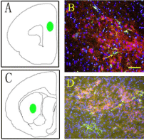
    Figure 1: Retro-grade tracing of meosocortical projection (A, B) and
mesostrial projection (C, D). A and C show the injection position in cerebral
cortex; Arrows in B and D show labeled subtype neurons of mesocortical and
mesostriatal projection, respectively.
One advantage of this tracing is that we can analize the physiological function with the brain slice patch. Dong et al in our research group labeled mesocortical subtype neurons and detected effect of aripiprazole, a new type of antipsychotic drug, on electrophysiological properties of labeled neurons with brain slice patch technique. Aripiprazole (10μM) reduced the frequency and amplitude of spontaneous action potentials (Figure 2).
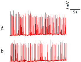
    Figure 2: Aripiprazole reduced the frequency and amplitude of spontaneous
action potentials. A, aripiprazole 10μM; B, control group.
Another advantage of microsphere retrobeads tracing is that the labeled cells can be collected through Fluorescence Activated Cell Sorting (FACS). And the RNA can be extracted from these pure subtype neurons, the further analysis, such as real time PCR and RNA-seq can be performed to explore the molecular mechanisms of specific neural projection.
Additionally, for the first time, we use microsphere retrobeads in peripheral nervous system. In our experiment, we cut sciatic nerve of rat, and the transected sciatic nerve was repared with muscle bridge technique. Eight weeks after transaction, retrobeads were hypodermicly injected into the foot of rat. Labeled neurons were detected in ipsilateral of spinal cord of rat (Figure 3).
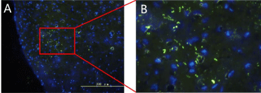
    Figure 3: Microsphere retrobeads show nerve regeneration.A, labeled
neurons in spinal cord; B, amplification of A.
In conclusion, microsphere retro-grade tracing provides a good technique for neural projection studies. We can not only labeled subtype neurons, but also can explore the underlying molecular mechanisms in cell specification. Moreover, we have already used this technique in peripheral nervous system for nerve regeneration detection.
References
- Tomassy GS, Lodato S, Gibson ZT, Arlotta P. Development and regeneration of projection neuron subtypes of the cerebral cortex. Sci Prog. 2010; 93: 151- 169.
- Zingg B, Hintiryan H, Gou L, Song MY, Bay M, Bienkowski MS, et al. Neural networks of the mouse neocortex. Cell. 2014; 156: 1096-1111.
- Katz LC, Iarovici DM. Green fluorescent latex microspheres: a new retrograde tracer. Neuroscience. 1990; 34: 511-520.