
Research Article
Austin J Anat. 2015; 2(3): 1038.
Imaging of Cranial Nerves
Tanrikulu L¹*, Hastreiter P¹, Buchfelder M¹, Jablawi F² and Naraghi R¹
¹Department of Neurosurgery, University of Erlangen- Nuremberg, Germany
²Department of Neurosurgery, RWTH Aachen University, Germany
*Corresponding author: Tanrikulu L, Department of Neurosurgery, RWTH Aachen University, Pauwelsstr, 30, Germany
Received: July 05, 2015; Accepted: September 29, 2015; Published: October 10, 2015
Abstract
Objective: In this study we present our experience in imaging and image processing of cranial nerve anatomy of the cranial nerves V-XI.
Material and Methods: We show the technique of imaging and image processing by high-resolution magnetic resonance imaging and threedimensional (3D) visualization by direct volume rendering. The cranial nerve anatomy is visualized referring the nerval root exit/entry zone, anatomical course within the cisterns of the posterior cranial fossa and the corresponding vasculature. Patients with trigeminal neuralgia, hemi facial spasm and gloss pharyngeal neuralgia were studied.
Results: With highly-resolute MR imaging and 3D visualization by direct volume rendering the anatomy of the cranial nerves V,VI,VII,VIII, IX,X and XI were delineated non-invasively in the pre- and intra operative setup of the surgical domain. There were no disturbances in anatomical visualization.
Conclusion: Cranial nerve imaging by highly resoluted MR imaging and 3D visualization by direct volume rendering gives the opportunity for non-invasive anatomical analysis of cranial nerve topography and corresponding vasculature and brainstem. This contributes for diagnostic and therapeutic decisions and for surgical-anatomical teaching.
Keywords: Cranial nerves; Imaging; Image processing; Neuroanatomy
Abbreviations
2D: two-Dimensional; 3D: three-Dimensional
Introduction
Cranial nerve anatomy and corresponding vasculature still remains as a challenging field for surgeons. There is a need for non-invasive anatomical visualization of the complex 3D topographical details of the cranial nerves and corresponding vessels especially in patients with trigeminal neuralgia, hemi facial spasm, glossopharyngeal neuralgia and for patients with tumors within the posterior cranial fossa such as acoustic neuromas, meningiomas or metastases. The goal of this article is to show our experience in imaging of cranial nerve anatomy and corresponding vessels. The question is whether we are able to transfer given two-Dimensional (2D) anatomical data into more interesting three-Dimensional (3D) delineations in order to get maximum anatomical information about the individual cranial nerve anatomy prior to surgery and at the intra operative surgical domain.
Material and Methods
From the years 2003 until 2006 we routinely examined and treated patients with cranial nerve rhizopathies such as trigeminal neuralgia (41 patients), hemi facial spasm (25 patients) and glossopharyngeal neuralgia (2 patients) [1-3]. All patients underwent high-resolution 1.5 Tesla magnetic resonance imaging described as constructive interference in the steady state (MRI-CISS, Figure 1) [4,5]. In MRICISS sequences the cisterns filled with Cerebro Spinal Fluid (CSF) are depicted with highest intensity and the containing anatomical structures such as the interesting cranial nerves and corresponding vessels are delineated hypo intensely (Figure 1). Therefore the individual three-dimensional anatomy of cranial nerves, vessels and the brainstem is visualized in each patient non-invasively and the position of the anatomical structures can be varied into all directions. The MRI-CISS sequences undergo segmentation (Figure 2) and subsequent 3D visualization by direct volume rendering (Figure 3) [4]. The result is a 3D object represent in the real, individual patient anatomy with topographical details of cranial nerves and surrounding structures as the brainstem and vessels (Figure 4). Our patients underwent micro vascular decompression after sub occipital, retrosigmoidal craniotomy as described by Jannetta [6].
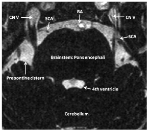
    Figure 1:  Slice of a high-resolution MR CISS (Constructive Interference in
the Steady State) image. Note: the CSF space (Prepontine Cistern, Fourth
Ventricle) are delineated highly hyper intense, so that the containing cranial
nerves (CN V: Trigeminal Nerves), vessels (BA: Basilar Artery, SCA:
Superior Cerebellar Artery) and the brainstem (Pons Encephali) are depicted
hypo intensely, so that a clear differentiation between these structures are
obtained.
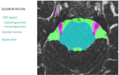
    Figure 2:  Procedure of Segmentation. The CSF spaces are segmented
semi automatically (green color) with the corresponding vessels, the cranial
nerves (purple color) and the brainstem (turquoise color) in each patient. After
this procedure the obtained segmented data is processed by direct volume
rendering where the segmented volumes CSF space, cranial nerves and
brainstem are transferred to opacity/transparency values and different colors.
The CSF space is transferred to a high opacity/low transparency volume, so
that the containing vessels are visualized and the brainstem and the cranial
nerves are transferred to high opacity/low transparency volumes.
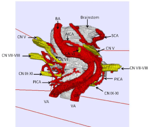
    Figure 3:  Resulting 3D visualization generated after direct volume rendering
from segmentation. Note: One can vary the position of the 3D object into each
direction in the space. The cranial nerves V-XI, the corresponding vessels
and the brainstem are visualized in a non-invasive way.
CN V: trigeminal nerve; CN VI: abducent nerve; CN VII: facial nerve; CN
VIII: vestibulocochlear nerve; CN IX: glossopharyngeal nerve; CN X: vagus
nerve; CN XI: accessory nerve; BA: Basilar Artery; SCA: Superior Cerebellar
Artery; AICA: Anterior Inferior Cerebellar artery; VA: Vertebral Artery; PICA:
Posterior Inferior Cerebellar Artery.
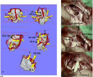
    Figure 4:  Real, individual 3D visualization in a patient with right sided
trigeminal neuralgia containing the interesting cranial nerves and
corresponding vessels. Figure 4a shows the variation of the position of 3D
visualization in correspondence to the surgical domain.
V: trigeminal nerve; VI: abducent nerve; VII: facial nerve; VIII: vestibulocochlear
nerve; IX: glossopharyngeal nerve; X: vagus nerve; XI: accessory nerve; BA:
basilar artery; VA: vertebral artery; SCA: Superior Cerebellar Artery; LA:
Labyrinthine Artery; AICA: Anterior Inferior Cerebellar Artery; f: Floccules
Cerebelli; mot.: motor division of trigeminal nerve; sens.: sensory division of
trigeminal nerve.
Results
We were able to visualize the cranial nerves (V, VI, VII, VIII, IX, X, and XI), the brainstem and the corresponding vasculature in all examined patients (100%) without any medical or technical disturbances. All patients underwent surgery (micro vascular decompression) and 3D visualization of the neurovascular conflicts were applied to the pre- and intra operative setup without any disturbance. The surgeon was able to see pre- and intraoperatively in addition to the available 2D MRI imaging the real, individual cranial nerve anatomy in 3D object with possible variation of its position into each direction, so that the coaxial view into the surgical domain could always be compared to the non-invasively reproduced 3D visualization of the patient.
Discussion
Two-dimensional images by magnetic resonance imaging do not contribute for the complex and detailed anatomical characteristics and relationships between the brainstem, cranial nerves and corresponding vessels in the posterior cranial fossa. With 3D visualization of the 2D imaged with direct volume rendering we were able to generate real, individual, anatomical representations of our examined patients prior to surgery. MRI CISS is surely superior in detailed anatomical imaging to “routine” MR sequences where the cranial nerve anatomy cannot be visualized sufficiently [5], because MRI-CISS allows a clear differentiation of nerves and vessels within the CSF spaces. One limitation of our method is the existence of pulsation artifacts induced by the pulsation of the basilar and vertebral arteries, while these artifacts could be deleted within the segmentation process by manual corrections in order to have a clear view over the surface of the brainstem. The 3D visualization represented a precondition for detailed anatomical analysis and understanding of volumetric data from the MR imaging. We were able to draw classifications of the neurovascular anatomy of facial-vestibulocochlear-nerve complex in patients with hemi facial spasm [4], because we routinely applied the technique of intra operative 3D visualization during micro vascular decompression in the operating theatre. This provided us a very advantegous possibility of anatomical information before and during surgery and contributed for anatomical and operative safety and clinical outcome of the patient.
Conclusion
With 3D visualization we were able to show the real, individual cranial nerve anatomy and corresponding vasculature and the brainstem in patients with neurovascular compression syndromes. This method of non-invasive anatomical exploration of the cranial nerves and vessels is very useful for topographical analysis and classifications of the variable vascular compression courses within the posterior cranial fossa. This method also contributes to diagnostic and therapeutic decisions in patient care and to surgical-anatomical teaching.
References
- Tanrikulu L, Hastreiter P, Richter G, Doerfler A, Naraghi R. Virtual neuroendoscopy: MRI-based three-dimensional visualization of the cranial nerves in the posterior cranial fossa. Br J Neurosurg. 2008; 22: 207-212.
- Naraghi R, Tanrikulu L, Troescher-Weber R, Bischoff B, Hecht M, Buchfelder M, et al. Classification of neurovascular compression in typical hemifacial spasm: three-dimensional visualization of the facial and the vestibulocochlear nerves. J Neurosurg. 2007; 107: 1154-1163.
- Tanrikulu L, Hastreiter P, Troescher-Weber R, Buchfelder M, Naraghi R. Intraoperative three-dimensional visualization in microvascular decompression. J Neurosurg. 2007; 107: 1137-1143.
- Hastreiter P, Naraghi R, Tomandl B, Bonk A, Fahlbusch R. Analysis and 3-dimensional visualization of neurovascular compression syndromes. Acad Radiol. 2003; 10: 1369-1379.
- Tomandl BF, Hastreiter P, Rezk-Salama C, Engel K, Ertl T, Huk WJ, et al. Local and remote visualization techniques for interactive direct volume rendering in neuroradiology. Radiographics. 2001; 21: 1561-1572.
- Jannetta PJ. Trigeminal neuralgia. Neurosurgery. 1986; 18: 677.