
Original Article
Austin J Anat. 2015;2(2): 1034.
Functional Adaptability of the Tunica Media of the Atriopulmonary Junction
Poonamjeet Kaur Loyal*, Kevin Ongeti, Pamela Mandela and Paul Odula
Department of Human Anatomy, University of Nairobi, Kenya
*Corresponding author: Poonamjeet Kaur Loyal,Department of Human Anatomy, University of Nairobi,School of Medicine, 30197-00100- Nairobi, Kenya
Received: March 03, 2015; Accepted: May 04, 2015; Published: May 04, 2015
Abstract
Introduction: The histoarchitecture of the tunica media of the Atriopulmonary Junction (APJ) has not been extensively studied and yet the tunica media is the backbone, which provides the structural strength for optimum function. This study therefore strives to unravel the structural components of the tunica media of the APJ.
Methods: The study design was a descriptive cross-sectional study. Histological studies were done on tissues from the APJ of 20 hearts using light microscopy. The proportion of elastic fibers in the tunica media of the APJ was determined using the point intercept method by dropping a grid in ImageJ software.
Results: The smooth muscle cells were concentric and more prominent in males extending the entire extent of the tunica media compared to females where they were confined to the intimal side of tunica media. The collagen bundles were mainly longitudinal. The elastic fibers showed a diverse pattern of organization and side and sex differences in orientation and quantity.
Conclusion: Gender and regional variations in the histology of the tunica media of the pulmonary veins suggest a mechanism for controlling hemodynamic forces at the APJ.
Keywords: Tunica media; Atriopulmonary junction
Introduction
The Atriopulmonary Junction (APJ) is an interface between the Left Atrium (LA) and the Pulmonary Veins (PVs) [1]. It is subjected to various forces including hemodynamic stress of the blood as it flows from the lungs to the left atrium; translational forces of the lung through the pulmonary veins; and retrograde pressure forces during systolic contractions of the left atrium [2]. The tunica media of a blood vessel consists of smooth muscle, elastic fiber and collagen bundles [1], but there is paucity of information regarding the histoarchitecture of the APJ. This study was therefore designed to investigate the functional adaptability of the tunica media of the APJ in the face of the volume of forces that it withstands.
Materials and Methods
Ethical approval was sought from the Kenyatta National Hospital- University of Nairobi Ethics and Research Committee before commencement of the study. Permission and requisite authority was obtained from the administration of Chiromo Funeral Parlor for acquiring histological tissues from autopsy specimens. Twenty hearts were used for histomorphology with 10 being from male and 10 from female subjects for a fairer representation. Since a large proportion of the victims of cardiovascular disease are from the adult age group [3], hearts from subjects ranging from 20-40 years were used as study specimens. All specimens were from individuals of black Kenyan descent.
Heart specimens from both sexes and of adult age group (>18 years) were included in this study. With the help of the pathologist, heart specimens with any observable pathology or congenital defects were excluded. Autopsy specimens that had stayed for more than 48 hours post mortem were also excluded from histological study.
Data collection procedure
At autopsy, with the help of the pathologist, a Y-incision was made from the acromio-clavicular joints to the xiphoid process. The thoracic cage was opened by incision of the costal cartilages. The sternum was then removed carefully and pericardium was incised longitudinally to expose the heart. The hearts were categorized according to sex and longitudinal and transverse sections of APJ were cut. This allowed regional histoarchitectural changes from the LA, APJ, and proximal part of PV to be studied. The tissues were fixed for a period of 72 hours in 10% formal saline after which they were dehydrated in ethyl alcohol of increasing concentrations, starting with 70% alcohol to absolute alcohol (1hour in each solution). Toluene was used as a clearing agent for 1.5 hours followed by wax impregnation for 12hours at 58°C. After tissues were embedded in freshly molted wax, 7 micron thick sections were prepared using a sledge microtome (Leica Model SM2400, Leica Microsystems, Nussloch GmbH, Germany). These were then floated in warm water at 37°C to enhance spreading upon which they will be fixed on slides using egg albumin. The slides were left to dry in an oven at 38°C for 12 hours. Dewaxing was done using xylene and the segments rehydrated using xylol (prepared from xylene and isopropyl column), absolute alcohol and descending concentrations of alcohol to 70% then running water. The sections were stained with Masson’s Trichrome (MT) stain to demonstrate connective tissue and myocardial sleeve organization and Weigert’s resorcin fuschsin with van Gieson counterstaining (WT) to demonstrate elastic fibers. Mounting of the cover slips onto specimens was done using D.P.X mountant (Unilab Kenya).The slides were examined with a light microscope (Leica model BME, Leica Microsystems, Nussloch GmbH, Germany) at x40, x100, x400 magnifications and the histological features were recorded on data sheets.The slides were photographed using a ZeissTM digital photomicroscope (Carl Zeiss AG, Oborkochen, Germany) at various magnifications and were then downloaded for further computer analysis. The photographs were edited using Photoscape Image Editor ™version 3.5. The proportion of elastic fibers in the tunica media of the APJ was determined using the point intercept method by dropping a grid in ImageJ software:
Results
Salient features of the tunica media of the APJ were flattened fusiform shaped smooth muscles amidst dense regular connective tissue consisting of collagen bundles and elastic fibers. The Smooth Muscle (SM) cells were concentric and more prominent in males (Figure 1) compared to females (Figure 2). Whereas the SM cells occupied almost the entire extent of the tunica media in the males (Figure 1A, 1B), in females they were confined to the intimal side of the tunica media (Figure 2A, 2B). The collagen bundles were mainly longitudinal but changes in orientation were observed towards the adventitial side of the tunica media as they became less densely packed (Figure 1, 2).
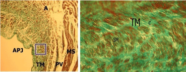
    Figure 1:  1A,1B (Inset): Photomicrograph of the longitudinal section of right
superior atriopulmonary junction in a 33 year old male. Note the densely
packed Collagen Bundles (C) with abundant Smooth Muscle cells (SM)
extending deep into the Tunica Media (TM) of the Atriopulmonary Junction
(APJ).
PV: Pulmonary Vein; MS: Myocardial Sleeves; C: Collagen bundles; A:
Atrium (MT X40, inset: x400).
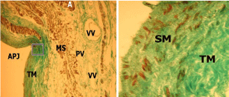
    Figure 2:  2A, 2B (Inset): Photomicrograph of a longitudinal section of the right
superior atriopulmonary junction in a 27 year old female. Note the Smooth
Muscle Cells (SMs) are oriented in varying directions and are confined to the
intimal zone of the Tunica Media (TM) in the Atriopulmonary Junction (APJ).
A: Atrium; PV: Pulmonary Vein; VV: Vasa Vasora; NV: Nervi Vasora; MS:
Myocardial Sleeve; C: Collagen bundles (MT X40, inset: x400).
The elastic fibers showed a diverse pattern of organization with both circular and longitudinal orientations observed. The elastic fibers displayed side and sex differences in orientation and quantity. The proportion of elastic fibers was greater in males than in females for both the anterior and posterior walls of the APJ (Table 1). Other differences were noted in orientation of the elastic fibers whereby males mainly had circular elastic lamellae in the posterior wall (Figure 3) and longitudinal elastic fibers (Figure 5) in the anterior wall. Females had a predominantly longitudinal disposition of the elastic fibers in both the posterior walls (Figure 4) and anterior walls (Figure 6). The elastic fibers in the tunica media also increased in thickness and became more filamentous towards the adventitia (Figure 3).
![]()
    Anterior    wall of junction (%)
    Posterior    wall of atriopulmonary junction (%)


    Male
    Female
    Male
    Female


    LS
    66.5
    32.2
    75.5
    51.6


    LI
    55.7
    41.7
    66.7
    30.5


    RS
    55.1
    27.0
    72.1
    48.6


    RI
    48.6
    27.3
    76.2
    33.3


    LS: Left    Superior Atriopulmonary Junction; LI: Left Inferior Atriopulmonary Junction;    RS: Right Superior Atriopulmonary Junction; RI: Right Inferior Atriopulmonary    Junction.
Table 1:  Percentage of elastic fibers in tunica media of the atriopulmonary
junction.
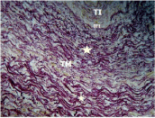
    Figure 3:  Photomicrograph of a transverse section of the posterior wall of the
left superior atriopulmonay junction of a 27-year-old male. Note the presence
of concentric elastic fibers (?) in the Tunica Media (TM), which increase in
fiber thickness and become longer filaments towards the adventitia.
TI: Tunica Intima; IEL: Internal Elastic Lamina (WE X400).
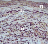
    Figure 4:  Photomicrograph of transverse section of the posterior wall of
the left superior atriopulmonary junction in a 25 year old female showing
preponderantly longitudinal elastic fibers (in the Tunica Media (TM).
TI: Tunica Intima; IEL: Internal Elastic Lamina (WE X400).
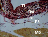
    Figure 5:  Photomicrograph of a transverse section of the anterior wall of the
left superior atriopulmonary junction in a 27 year old male showing relatively
fewer elastic fibers in the Tunica Media (TM) with predominantly longitudinal
dispositions.
TI: Tunica Intima; EEL: External Elastic Lamina; PS: Perimuscular Sheath;
MS: Myocardial Sleeves (WE X400).
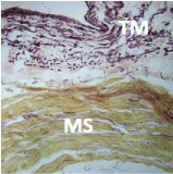
    Figure 6:  Photomicrograph of transverse section of the anterior wall of the
Left superior atriopulmonary junction in a 25 year old female with elastic fibers
in the Tunica Media (TM) oriented in varying directions but a predominance
of longitudinal arrangement.
TM: Tunica Media; MS: Myocardial Sleeve (WE X400).
Discussion
In tandem with previous reports [2], the tunica media was observed to have concentric smooth muscle fibers interspersed in dense connective tissue consisting of collagen bundles and elastic fibers. An additional finding in the present study was the existence of gender and side differences in the fiber orientation and density.
A circular pattern of orientation of smooth muscle cells was observed, and noted to be similar to that in the aorta. This supports the theory of pulsatile flow of blood in the PV, previously considered as passive conduits [4]. Contraction of these cells provides the propulsive force for the maintenance of a constant pulsatile stream of blood from the lungs to the atria. This is comparable to the pulsatile nature of blood flow in the aorta that has been attributed to the presence of smooth muscle cells [5]. Furthermore, the involvement of these smooth muscle cells in instigating myogenic response at the junction is important for the local regulations of blood flow and generation of basal vascular tone cannot be overlooked [6].
There were sex variations noted in the content of the smooth muscle cells in the tunica media. The more densely packed smooth muscles in the tunica media in males than in females may be a result of differential exposure to testosterone in utero [7]. Testosterone has been linked to inducing vascular smooth muscle migration and its relative abundance in utero for males, which is also maintained postnatally, may explain the densely packed nature of smooth muscle cells in the male specimens compared to female [7].
The abundant presence of elastic fibers in the tunica media accords unique physiological properties to particular anatomical sites where they are found. Elastic fibers possess rubber like properties; they can stretch easily and rapidly, and can return to their prestressed state with minimal loss of energy, thereby providing elasticity and stretchability to tissue [8]. The elastic fibers of the APJ are comparable to those found in elastic arteries. It is, therefore, not surprising that the pulmonary venous system should contribute upto 60% of the compliance of the pulmonary vascular bed [9]. Previous studies done on the aorta have attributed the elastic lamellae to be involved in the windkessel function whereby during systole, a fraction of the cardiac output is accommodated to be released in diastole when the elastic fibers relax [10]. Although reports of such a phenomenon are largely unreported for other vessels, the APJ has been linked to capacitance functions which seem comparable to the windkessel mechanism of the aorta. Past observations seem to support this view, whereby an increase in the left atrial pressures were seen to cause a decrease in the resistance of the pulmonary venous system primarily due to distension forces contributed by elastic fibers [11]. The reverse is noted when the left atrial pressure decrease, and the elastic recoil facilitates the blood flow to the LA.
Another possible function of the elastic fibers may be to act as shock pads at the APJ. Lung capillary outflow is largely protected from the pressure events that occur in the left atrium [12]. This may be as a result of the elastic fibers absorbing the retrograde pressure pulsations originating during systolic contractions of the left atrium. Presence of circular elastic fibers at junctions is commonly reported; where they instill resilience and compliance as is the requirement for structures subjected to incessant distension and contraction [13]. These circular elastic fibers were more common on the posterior wall compared to the anterior wall, further being greater in the male specimens. This infers that the posterior wall is the main component in maintaining the capacitance function and this function is more enhanced in males. The higher elastic content of the junction in males may be a functional adaptation in response to the greater hemodynamic stresses owing to the higher blood pressure in males [14]. The elastic fibers in females and in particular the anterior wall of males showed a predominantly longitudinal disposition, which may allow for a change in the length of the PVs corresponding to the lung movements during respiration. This may be the reason why the PVs are not torn off from the heart at the APJ, when subjected to pull forces during inspiration. The predominance of the longitudinal arrangement in females may be a function of the differential breathing patterns adopted by the different sexes. It is known that males exhibit predominantly abdominothoracic breathing pattern, whereas females are seen to mainly have a diaphragmatic or thoracoabdominal pattern [15]. The more rapid action of the rib cage muscles in thoracoabdominal breathing [16] facilitates greater lung movements in the vertical plane in females, predisposing them to more shearing forces at the APJ. The predominantly longitudinal elastic fiber arrangement in females may therefore be an adopted characteristic to allow for a change in length corresponding to their lung movements during respiration.
Collagen seems to be the main load bearing component of the junction responsible for structural integrity and tensile strength [17]. The compact and regular arrangement of collagen fibers in the tunica media serves to increase the ability of the junction to better withstand hemodynamic stresses. The present study demonstrated a concurrent presence of elastic and collagen fibers in the tunica media of the APJ suggesting integrative structural and functional interactions. Previous studies have reported on the synergistic functions of elastic and collagen fibers whereby low transmural pressure is mainly borne by elastic fibers, engaging collagen fibers only as transmural pressures rise [18]. This may augment functional adaptability of the junction to hemodynamic stresses by preventing one fiber system from being overworked by bearing all the strain forces. It is thus plausible that the biomechanical properties of the APJ will be discordant with Hooke’s Law since the more the junction is subjected to stretch forces the more it will tend to resist. This is supported by Apter (1967), who attributed the resultant non-linear compliance of blood vessels, to the relative indistensibility of the collagen fibers.
Conclusion
In conclusion, the tunica media of the atriopulmonary junction is well endowed structurally to withstand the myriad of forces attacking the interface. Gender and regional variations in the histology of the tunica media of the pulmonary veins suggest a mechanism for controlling hemodynamic forces at the atriopulmonary junction.
Acknowledgement
Poonamjeet Loyal: Was involved in concept/design, acquisition of data, data analysis/interpretation, drafting of the manuscript, critical revision of the manuscript and approval of the article.
Kevin Ongeti: Involved in acquisition of data, data analysis/ interpretation, drafting of the manuscript, critical revision of the manuscript and approval of the article.
Pamela Mandela: concept/design, data analysis/interpretation, drafting of the manuscript, critical revision of the manuscript and approval of the article.
Paul Odula: concept/design, acquisition of data, drafting of the manuscript, critical revision of the manuscript and approval of the article.
References
- Standring S, Ellis H, Healy JC (2004) Thorax. In: Gray’s Anatomy 39th edition. Elsevier Churchill Livingstone, London. 1027.
- Steiner I, Hajkova P, Kvasnicka J, Kholova I. Myocardial sleeves of pulmonary veins and atrial fibrillation: a postmortem histopathological study of 100 subjects. Virchows Arch. 2006; 449: 88-95.
- Habibzadeh F, Yadollahie M, Roshanipoor M, Haghighi AB. Prevalence of atrial fibrillation in a primary health care centre in Fars province, Islamic Republic of Iran. East Mediterranean Health Journal. 2004; 10:147-151.
- Smiseth OA, Thompson CR, Lohavanichbutr K, Ling H, Abel JG, Miyagishima RT, et al. The pulmonary venous systolic flow pulse�its origin and relationship to left atrial pressure. J Am Coll Cardiol. 1999; 34: 802-809.
- Orsi AM, Stefanini MA, Crocci AJ, Simoes K, Ribeiro AA. Some segmental features on the structure of the aortic wall of the dog. Anat Histol Embryol. 2004; 33:131-134.
- Meininger GA, Davis MJ. Cellular mechanisms involved in the vascular myogenic response. Am J Physiol.1992; 263: H647-H659.
- Chignalia AZ, Schuldt EZ, Camargo LL, Montezano AC, Callera GE, Laurindo FR, et al. Testosterone induces vascular smooth muscle cell migration by NADPH oxidase and c-Src-dependent pathways. Hypertension. 2012; 59: 1263-1271.
- Davidson JM, Zang MC, Zoia O, Giro MG. Regulation of elastin synthesis in pathological states. Ciba Found Symp. 1995; 192: 81-99.
- Shoukas AA. Pressure-flow and pressure-volume relations in the active pulmonary vascular bed of the dog determined by two-port analysis. Circ Res. 1975; 37: 809-818.
- Robert L, Jacob MP, Fulop T. Elastin in blood vessels. Ciba Found Symp. 1995; 192: 286-303.
- Kuramoto K, Rodbard S. Effects of blood flow and left atrial pressure on pulmonary venous resistance. Circ Res. 1962; 11: 240-246.
- Lee GJ. Regulation of the pulmonary circulation. Brit Heart J. 1971; 33: 15-26.
- Reisdoerfer G, Pereira KF, Chopard RP. Fibroelastic components of the vesicourethral junction of rats in the aging process. Int J Morphol. 2011; 29: 214-220.
- Wallin BG, Hart EC, Wehrwein EA, Charkoudian N, Joyner MJ. Relationship between breathing and cardiovascular function at rest: sex-related differences. Acta Physiol (Oxf). 2010; 200: 193-200.
- Wang CS, Josenhans WT. Contribution of diaphragmatic/abdominal displacement to ventilation in supine man. J Appl Physiol. 1971; 31: 576-580.
- Sharp JT, Goldberg NB, Druz WS, Danon J. Relative contributions of rib cage and abdomen to breathing in normal subject. J Appl Physiol. 1975; 39: 608-618.
- Harkness ML, Harkness RD, McDonald DA. The collagen and elastin content of the arterial wall in the dog. Proc Roy Soc London. 1957; 146: 541-551.
- Apter JT. Correlation of visco-elastic properties with microscopic structure of large arteries: thermal responses of collagen, elastin, smooth muscle, and intact arteries. Circ Res. 1967; 21: 901-918.