Department of Pathology, Anatomy and Cell Biology, Thomas Jefferson University, USA
*Corresponding author: Zhang G, Department of Pathology, Anatomy and Cell Biology, Thomas Jefferson University, 1020 Locust Street, Room 263 Jefferson Alumni Hall, Philadelphia, PA 19107. USA.
Received: April 20, 2014; Accepted:November 22, 2014; Published: December 01, 2014
Citation: Zhang G and Fenderson BA. Pathology Encountered During Cadaver Dissection Provides an Opportunity for Integrated Learning and Critical Thinking. Austin J Anat. 2014;1(5): 1027. ISSN:2381-8921
Cadaver dissection engages medical students in active learning, critical thinking, and problem solving. During dissection, students at Sidney Kimmel Medical College are encouraged to document pathologic findings in their cadavers and discuss the findings with their peers. Here, we describe two cases that provided opportunities for integrating anatomy with pathology and clinical medicine. The first case was a benign ovarian neoplasm. This large pelvic mass displaced the urinary bladder and uterus, and compressed the ureters and rectum. Students discussed clinical complications of this pelvic mass and reviewed pathologic findings that differentiate benign from malignant neoplasms. The second case was breast cancer with widespread metastases and pulmonary atelectasis. Students examined the histopathology of breast cancer and discussed mechanisms of tumor metastasis. Pathologic findings and evidence of past surgical procedures are frequently encountered during cadaver dissection. These findings provide a valuable opportunity for integrating the preclinical basic sciences and stimulating the development of critical thinking skills.
Keywords: Medical education; Cadaver dissection; Pathology; Ovarian adenoma; Breast adenocarcinoma
A: Atelectasis; B: Urinary bladder; C: Cecum; D: Diaphragm; H: Heart; L: Liver; O: Ovary; RL: Right lung; S: Sigmoid colon; T: Uterine tube; U: Uterus; Ur: Ureter
The goals of preclinical basic science education are to provide an integrated scientific foundation for medical practice. Students learn to critically evaluate clinicopathologic findings and generate informed decisions for the benefit of patient care. The traditional medical curriculum consists of separate courses in human anatomy, biochemistry, histology, physiology, neuroscience, and pathology. Although most medical students and allied health professionals form the appropriate connections that are required for clinical problem solving [1,2], most medical educators are keenly aware of the need for integration of the preclinical basic science curriculum [3,4]. Moreover, medical educators are encouraged to adopt classroom and laboratory learning activities that promote active learning and critical thinking [5].
Sidney Kimmel Medical College of Thomas Jefferson University follows a traditional formula for gross anatomy instruction based on overview lectures and cadaver dissection [6]. Over the years, we have attempted to improve our curriculum by creating a “toolkit” of active learning experiences for our students. Active learning encourages collaboration and independent study, involves learners in the educational process, highlights clinical relevance, illustrates scientific and clinical reasoning, and generally celebrates the joy of learning. To promote active learning, our anatomy students are encouraged to document pathologic findings observed in their cadavers. Here, we describe two cases that provided rich opportunities for integrating anatomy with pathology and clinical medicine. These cases promoted student-faculty dialogue and fostered the development of critical thinking skills.
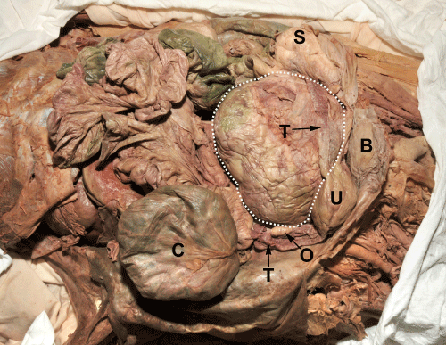
Dissection of an elderly female cadaver revealed a large tumor mass filling the entire true pelvis (Figure 1). The tumor displayed a smooth, connective tissue capsule. The superior aspect of the tumor protruded into the greater pelvis and was adherent to a portion of the small intestine. The urinary bladder and uterus were displaced anteriorly and superiorly into the greater pelvis. The uterus deviated from the midline and was located on the right lateral border of the urinary bladder. A loop of the sigmoid colon was pushed into the abdominal cavity on the left side. The cecum was greatly extended and filled with gas. The left uterine tube appeared to have been stretched and extended; it lacked normal tubular morphology and was attached to the left superior margin of the tumor. The right uterine tube and ovary appeared normal.
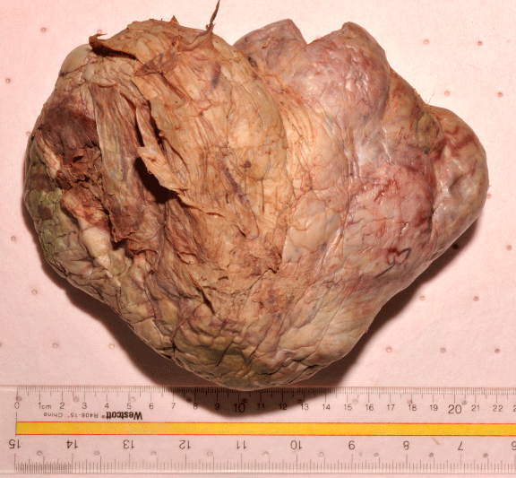
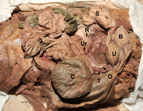
The isolated ovarian tumor was encapsulated and measured 22×15×15 cm (Figure 2). Incision of the tumor revealed small cystic spaces, suggesting a diagnosis of “ovarian cystadenoma”. Examination of the pelvis with the tumor removed showed that the rectum was compressed to form a thin flat muscular flap (Figure 3). The ureters were compressed against the posterior pelvic wall. Both renal pelves were dilated and their walls were thin (hydroureter). Sections through the kidneys revealed dilated major and minor calyces (hydronephrosis).
Based on these anatomic and pathologic findings, the faculty asked the students questions to promote group discussion: Why is the cecum extended and filled with gas? How did growth of the tumor affect the function of the gastrointestinal tract? Did compression of the ureters impair renal function? Are there other structures that could have been compressed and impaired by this pelvic mass? What clinical symptoms might the patient have experienced prior to death? The students reviewed normal pelvic anatomy and observed the effects of this tumor on those relationships. Because the tumor pressed against posterior and lateral walls of the pelvis, the students hypothesized that obturator nerves, sacral plexuses, and branches of the internal iliac arteries might have been compressed by this large ovarian tumor, resulting in associated neurovascular lesions. Tumor encapsulation was discussed as a characteristic feature of benign neoplasms.
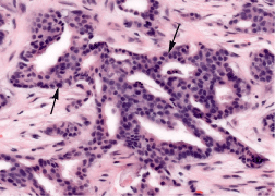
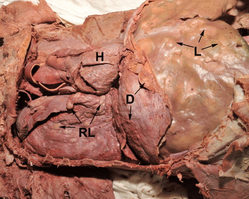
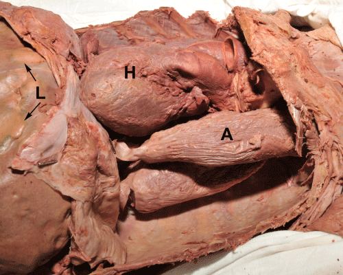
Dissection of the pectoral region of an elderly female cadaver with signs of cachexia revealed a unilateral mastectomy with silicone breast implant. Further exploration of the thorax and abdomen revealed widespread tumor metastases. Cannonball metastases were visible on the right lung and throughout the liver (Figure 4). Numerous tumor nodules were also evident on the costal and diaphragmatic pleura. Mediastinal lymph nodes were involved with malignant cells. Consistent with cancer cachexia in this patient, the epicardium, greater omentum, and mesentery of the gut were devoid of adipose connective tissue. Collapse of the left lung (pulmonary atelectasis) was apparently caused by tumor obstruction of the left main stem bronchus (Figure 5). Evidence of malignant disease was apparent throughout the abdomen and pelvis, including pancreas, intestines, para-aortic lymph nodes, kidney, and uterus (results not shown). Malignant cells were not detected in the spleen. Histopathologic features of in situ and invasive cancer of the breast were reviewed using digital microscope slides obtained from the Jefferson Digital Slide Box (Figure 6).
Based on these pathologic findings, the faculty asked the students questions to promote group discussion and stimulate critical thinking: What are the risk factors for breast cancer? Discuss the TNM tumor staging system. Discuss differences between tumor staging and grading? List key steps in tumor metastasis. Discuss routes of tumor metastasis in this patient. Discuss mechanisms of cancer cachexia. What is the mechanism that resulted in pulmonary atelectasis? What other conditions can cause collapse of the lung?
Pathologic findings encountered during cadaver dissection provide an excellent opportunity for integrating the preclinical basic sciences and encouraging critical thinking. The cases reported here illustrate complications of benign and malignant neoplasms, and emphasize the importance of gross and microscopic anatomy for understanding the pathologic basis of disease. Pathologic findings encountered during cadaver dissection encourage students to make careful observations, formulate good questions, and draw conclusions based on evidence. The students become excited and engaged in learning. In the future, we plan to examine test scores and student surveys to demonstrate improvements in problem solving.
In this connection, a crucial aspect of clinical problem solving is the ability of the beginning student, and the practitioner, to ask informed questions [5,7]. Students are erroneously led to believe that experts have correct answers, whereas students have mere questions. On the contrary, questions are golden. They provide a necessary first step in the process of collecting data and developing a differential diagnosis. Pathologic findings encountered during cadaver dissection provide a welcome opportunity for educators to solicit questions and engage students in active learning. We believe that the ability of students to formulate thoughtful questions is a foundational skill that will enable them to make well-informed decisions for the benefit of patient care.
We wish thank the Humanity Gifts Registry and the many families who donate bodies of their loved ones for training the next generation of medical caregivers. Humanity Gifts Registry is a non-profit agency of the Commonwealth of Pennsylvania concerned primarily with the receipt and distribution of bodies donated to all medical and dental schools in the state for teaching purposes. Histopathology of breast cancer (Figure 6) was obtained from a digital microscope slide provided for educational use by Dr. Emanuel Rubin, Sidney Kimmel Medical College. We are proud to recognize the Thomas Jefferson University, Doctor of Physical Therapy class of 2016, for their professionalism and enthusiasm for learning.
A massive ovarian tumor occupies the entire lesser pelvis. The outline of the tumor is indicated by the dotted line.
Abbreviations: B: Urinary bladder; C: Cecum; O: Right ovary; S: Sigmoid colon; T: Uterine tube; U: Uterus
The isolated ovarian tumor exhibits a smooth connective tissue capsule. The 22×15×15 cm mass appears to be a benign epithelial neoplasm, most likely cystadenoma.
The pelvic cavity is examined with the ovarian tumor removed.
Abbreviations: B: Urinary bladder; C: Cecum; O: Right ovary; R: Rectum; S: Sigmoid colon; T: Uterine tube; U: Uterus; Ur: ureter
Nodules of metastatic breast cancer are observed in the lung, diaphragm, and liver (arrows). Abbreviations: D: Diaphragm; H: Heart; L: liver; RL: Right lung.
Both lobes of the left lung exhibit atelectasis, owing to obstruction of the main stem bronchus. The liver exhibits metastatic tumor colonies (arrows). Abbreviations: A: Atelectasis; H: Heart; L: Liver
Microscopic examination of invasive ductal carcinoma of breast from another patient reveals nests of epithelial cancer cells (arrows) that have elicited a conspicuous desmoplastic reaction.
