Case Report
Austin J Clin Case Rep. 2014;1(3): 1014.
Wrist Clonus and Oculomotor Apraxia in Corticobasal Degeneration
Jain Rajendra Singh1, Kumar Sunil1*, Nagpal Kadam1, Aggarwal Rakesh1 and Gupta Pankaj1
1Department of Neurology, SMS Medical College, India
*Corresponding author: Kumar Sunil, Department of Neurology, SMS Medical College, Jaipur, Rajasthan, India
Received: May 26, 2014; Accepted: June 10, 2014; Published: June 14, 2014
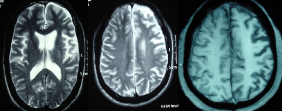
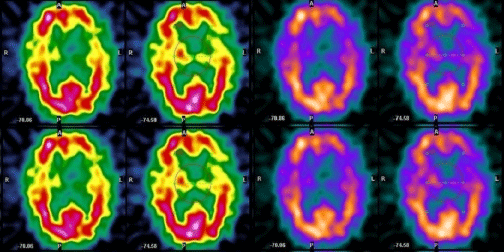
A 50 years old male presented with two years history of insidious onset gradually progressive asymmetric pyramidal and extrapyramidal features (right >left) with dysarthria and cognitive decline. His minimental status examination was 12/30 and detailed neuropsychological examination revealed dysfunction of dominant frontal and parietal lobes. Additionally, two uncommon findings oculomotor apraxia and wrist clonus (Video 1) were observed. The possibilities of corticobasal degeneration, bilateral opercular syndrome, and frontotemporal degeneration, nonfluent variant of primary progressive aphasia and frontal variant of Alzheimer disease were thought. MRI brain showed left fronto-parietal lobe atrophy (Figure 1) and SPECT revealed hypoperfusion in left parietal, bilateral basal ganglia and thalami but preserved perfusion in bilateral frontal, temporal and occipital lobes (Figure 2), consistent with diagnosis of corticobasal degeneration. To the best of our knowledge, wrist clonus has not been described in corticobasal degeneration so far. Our case was unique in view of unusual clinical signs of wrist clonus and oculomotor apraxia.